Compartment Syndromes and Volkmann Contracture

Chapter 74 Compartment Syndromes and Volkmann Contracture


Definition and History


Compartment syndrome is a condition in which the circulation within a closed compartment is compromised by an increase in pressure within the compartment, causing necrosis of muscles and nerves and eventually of the skin because of excessive swelling. Volkmann ischemic contracture is a sequela of untreated or inadequately treated compartment syndrome in which necrotic muscle and nerve tissue has been replaced with fibrous tissue.


In the upper extremity, compartment syndrome is most common in the forearm. The intrinsic muscle compartments of the hand also may be involved, and compartment syndrome of the upper arm has been reported.


In 1881, Volkmann stated in his classic paper that the paralytic contractures that could develop in only a few hours after injury were caused by arterial insufficiency or ischemia of the muscles. He suggested that tight bandages were the cause of the vascular insufficiency. This concept of extrinsic pressure as the primary cause of paralytic contracture persisted for some time in the English literature. In 1909, Thomas studied 107 paralytic contractures and found that some followed severe contusions of the forearm without fractures or splints or bandages applied to the limb. The idea was established that extrinsic pressure was not the sole cause of the ischemia. In 1914, Murphy reported that hemorrhage and effusion into the muscles could cause internal pressures to increase within the unyielding deep fascial compartments of the forearm with subsequent obstruction of the venous return. In 1928, Jones concluded that Volkmann contracture could be caused by pressure from within, from without, or from both. Eichler and Lipscomb outlined the early technique of fasciotomy as the primary surgical treatment.


Anatomy


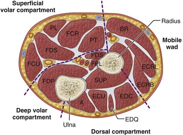
Four interconnected compartments of the forearm are recognized (Fig. 74-1): (1) the superficial volar compartment, (2) the deep volar compartment, (3) the dorsal compartment, (4) and the compartment containing the mobile wad of Henry (brachioradialis and extensor carpi radialis longus and brevis). The volar compartments are most commonly involved, but the dorsal and mobile wad compartments can be involved alone or in addition to the volar compartments. Clinically differentiating isolated or combined involvement of the deep and superficial volar compartments usually is difficult; however, the deep volar compartment (flexor digitorum profundus, flexor pollicis longus, and pronator quadratus) may be solely involved.


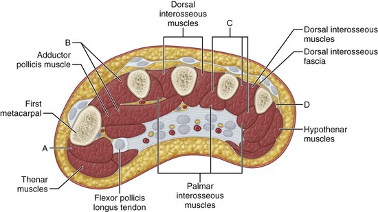
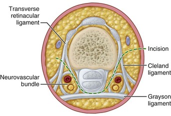
In the hand, each interosseous muscle is surrounded by a tough investing fascial layer, each making an individual compartment as shown by the injection dissections of Halpern and Mochizuki. The adductor pollicis muscle and the thenar and hypothenar muscles form three separate compartments (Fig. 74-2). The neurovascular bundles of each digit also are compartmentalized by fascial layers, making them vulnerable to excessive swelling (Fig. 74-3).


Etiology


Numerous injuries have been shown to result in compartment syndrome, including crush injuries, prolonged external compression, internal bleeding (especially after injury in patients with hemophilia), fractures, excessive exercise, burns, snake bites, and intraarterial injections of drugs or sclerosing agents. Infections also have been noted to increase pressures within compartments.


Elliott and Johnstone found that 18% of forearm compartment syndromes were caused by fractures and 23% were caused by soft tissue injuries without fractures. Although isolated distal radial fractures rarely were associated with compartment syndrome (0.3%), an ipsilateral elbow injury resulted in forearm compartment syndrome in 15% of patients. Historically, supracondylar humeral fractures were most frequently associated with forearm compartment syndrome in children; however, Grottkau et al. found that forearm fractures were actually more commonly associated (74% vs. 15%). Compartment syndrome after intramedullary fixation of forearm fractures in children also has been reported.


Acute compartment syndrome of the intrinsic muscles of the hand, resulting in contracture or necrosis of the muscle bellies, as in the larger muscles in the forearm, can occur after compression injuries of the hand without fracture. Compartment syndrome has been noted in neonates from intrauterine malposition or strangulation of the extremity by the umbilical cord.


Chronic exertional compartment syndromes of the first dorsal interosseous muscle and of the volar forearm also have been reported and occur most frequently in motorcyclists.


Direct trauma, crushing of the upper arm, shoulder dislocation, avulsion of the triceps muscle, pneumatic tourniquet use, and arteriography all have been reported as causes of compartment syndrome. Intravenous regional anesthesia also has been implicated as a cause when hypertonic saline is used to dilute an anesthetic.


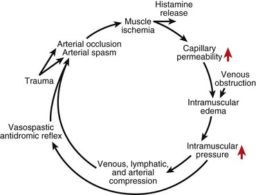
Any situation that causes a decrease in compartment size or increase in compartment pressure can initiate compartment syndrome. As the intracompartmental pressure increases, capillary blood perfusion is reduced to a level that cannot maintain tissue viability. The increase in interstitial pressure overcomes the intravascular pressure of the small vessels and capillaries, causing the walls to collapse and impeding local blood flow. In a canine model, muscle necrosis was shown to occur with a rise in pressure to within 20 mm below diastolic pressure. The local tissue ischemia leads to local edema, which increases intracompartmental pressure. This cycle of increasing muscle ischemia was depicted by Eaton and Green as shown in Figure 74-4.


The tolerance of tissue to prolonged ischemia varies according to the type of tissue. Functional impairment in muscles has been demonstrated after 2 to 4 hours of ischemia, and irreversible functional loss occurs after 4 to 12 hours. Nerve tissue shows abnormal function after 30 minutes of ischemia, with irreversible functional loss after 12 to 24 hours.


Diagnosis


A crush injury or fracture of the forearm or elbow, especially in the supracondylar area of the humerus, should raise suspicion that a forearm compartment syndrome may develop. Early diagnosis of impending ischemia is essential because irreversible damage can occur quickly. Increasing pain out of proportion to the injury that is worsened with passive stretch of the involved muscles is an early indication that a compartment syndrome is developing. The volar and or dorsal forearm is tender and tense with swelling, and sensibility of the fingertips may be diminished. Two-point discrimination and 256 cycles/vibratory testing can be helpful in determining nerve ischemia. Loss of the radial and or ulnar pulse usually develops later unless there is direct arterial injury. The diagnosis of a compartment syndrome of an individual interosseous muscle can be difficult. The hand is swollen and tense, and the fingers are held almost rigid in a partially flexed position with the wrist in neutral. Any passive movement of the fingers that causes metacarpophalangeal joint extension usually causes considerable pain. The adductor compartment of the thumb can be tested by pulling the thumb into palmar abduction and stretching the adductor muscle. The thenar muscles rarely are involved. Diagnosis in the obtunded and pediatric patient is more difficult. Compartment syndrome in a neonate may manifest as a sentinel bullous or ulcerative skin lesion usually over the dorsum of the forearm, wrist, or hand.


When a compartment syndrome is suspected and necessary equipment is available, compartment pressures should be obtained to confirm the diagnosis. Compartment pressures over 30 mm Hg or within 20 mm Hg of the diastolic pressure are indicative of compartment syndrome. All involved compartments should be measured and the results interpreted with regard to the overall clinical picture. Measurement may be obtained in the forearm from the superficial and deep volar compartments as well as the mobile wad and dorsal compartments. Hand measurements may be obtained from the thenar, hypothenar, adductor pollicis, and interosseous muscle. Digital pressures are not routinely obtained.


In 1975, Whitesides et al. described a technique of measuring compartment pressures using an 18-gauge needle, saline syringe, three-way stopcock, and a mercury manometer; however, currently a handheld pressure monitoring device or an arterial line monitoring system connected to either a straight needle, a side-port needle, or slit catheter is preferred. Boody compared the intracompartmental pressure monitoring system, an arterial line manometer, and the Whitesides apparatus each with a straight needle, a side-port needle, and a slit catheter and found that the arterial line manometer with the slit catheter was the most accurate technique. The handheld pressure monitoring system also was found to be accurate. Side-port needles and slit catheters were more accurate, whereas straight needles tended to overestimate the pressure. We most commonly use the handheld pressure monitoring device to determine intracompartmental pressures. The arterial line monitoring system is useful if continuous monitoring is desired.


To use the handheld pressure monitoring device (Stryker), the needle is placed firmly onto the chamber stem, a prefilled syringe is placed into the remaining chamber stem, and the chamber is firmly seated into the device. The needle is held at 45 degrees from horizontal and the system is purged of excess air. When the unit is turned on, the reading should be 0 to 9 mm Hg. To calibrate the system, the zero button should be pressed and the display should read 00. The needle is then inserted into the desired compartment, and no more than 0.3 mL is injected. The device then displays the pressure of the compartment.


Measuring Compartment Pressures in the Forearm and Hand Using a Hand-Held Monitoring Device


Technique 74-1


(LIPSCHITZ AND LIFCHEZ)


Management


Acute Compartment Syndrome of the Forearm


Impending tissue ischemia may be considered when the tissue pressure reaches between 30 mm Hg and 20 mm Hg below the diastolic blood pressure. A higher pressure is a strong indication that fasciotomy should be recommended. In a hypotensive patient, the acceptable pressure is lower. Fasciotomy should be performed in (1) normotensive patients with positive clinical findings and compartment pressures of greater than 30 mm Hg and when the duration of the increased pressure is unknown or thought to be longer than 8 hours; (2) uncooperative or unconscious patients with a compartment pressure greater than 30 mm Hg; and (3) patients with low blood pressure and a compartment pressure greater than 20 mm Hg. As a general rule, when in doubt, the compartment should be released. If it proves later to have been unnecessary, only a scar is the result. If a fasciotomy should have been done but was not, however, loss of muscle tissue and worse may result. A delay in diagnosis was the most important determining factor of poor outcome in one study. Compartment pressure should be monitored in young patients with injury to the forearm diaphysis or distal radius or in patients with significant soft tissue injury with a bleeding diathesis. Normal function was regained in 68% of patients in one study when fasciotomy was performed within 12 hours from the onset of compartment syndrome. When performing a volar fasciotomy, a volar curvilinear incision is used that allows release of the lacertus fibrosus proximally and the carpal tunnel distally. The interval between the flexor carpi ulnaris and the flexor digitorum sublimis is used for release of deep and superficial compartments. The dorsal forearm fascia is released through the interval between the extensor carpi radialis brevis and the extensor digitorum communis.


Forearm Fasciotomy and Arterial Exploration


Technique 74-2


image 
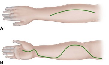
For the volar fasciotomy, make a curvilinear incision similar to McConnell’s combined exposure of the median and ulnar nerve neurovascular bundles as described by Henry (Fig. 74-5). Make an anterior curvilinear incision medial to the biceps tendon, crossing the elbow flexion crease at an angle. Carry the incision distally into the palm to allow for a carpal tunnel release, but avoid crossing the wrist flexion crease at a right angle.


image Divide the lacertus fibrosus proximally, and evacuate any hematoma.


image In patients with suspected brachial artery injury, expose the brachial artery and determine whether there is a free blood flow. If the flow is unsatisfactory, remove the adventitia to expose any underlying clot, spasm, or intimal tear. Resect the adventitia if necessary, and anastomose or graft the artery.


image Release the superficial volar compartment throughout its length with open scissors, freeing the fascia over the superficial compartment muscles.


image Identify the flexor carpi ulnaris, and retract it with its underlying ulnar neurovascular bundle medially, and retract the flexor digitorum superficialis and median nerve laterally to expose the flexor digitorum profundus in its deep compartment. Check to see if its overlying fascia or epimysium is tight, and incise it longitudinally.


image If the muscle is gray or dusky, the prognosis for recovery may be poor; however, the muscle may still be viable and should be allowed to perfuse.


image Continue the dissection distally by incising the transverse carpal ligament along the ulnar border of the palmaris longus tendon and median nerve.


image In cases of median nerve palsy or paresthesias, observe the median nerve along the entire zone of injury to ensure that it is not severed, contused, or entrapped between the ulnar and humeral head of the pronator teres. If it is, a partial pronator tenotomy is necessary.


image In a patient with a supracondylar fracture, reduce the fracture, pin it with Kirschner wires, and control the bleeding.


image Do not close the skin at this time; anticipate secondary closure later.


image If the median nerve is exposed within the distal forearm, suture the distal radial-based forearm flap loosely over the nerve.


image Check the dorsal compartments clinically, or repeat the pressure measurements. Usually, the volar fasciotomy decompresses the dorsal musculature sufficiently, but if involvement of the dorsal compartments is still suspected, release them also.


image Make the incision distal to the lateral epicondyle between the extensor digitorum communis and extensor carpi radialis brevis, extending approximately 10 cm distally. Gently undermine the subcutaneous tissue, and release the fascia overlying the mobile wad of Henry and the extensor retinaculum.


image Apply a sterile moist dressing and a long-arm splint. The elbow should not be left flexed beyond 90 degrees.


Jun 5, 2016 | Posted by in ORTHOPEDIC | Comments Off on Compartment Syndromes and Volkmann Contracture

Full access? Get Clinical Tree

Get Clinical Tree app for offline access