Arthrodesis of the Hip

Chapter 5 Arthrodesis of the Hip


The first successful arthrodesis of the hip was reported in 1894, and since then a variety of techniques for hip arthrodesis have been described. Early procedures were intraarticular and required extensive postoperative immobilization. Later, extraarticular arthrodesis using iliofemoral bone grafts was introduced, as well as subtrochanteric osteotomy to improve fusion rates by removing the disruptive force of the long femoral lever arm on the hip. Internal fixation was introduced by Watson-Jones and others in the 1930s and improved upon by Charnley; however, these early methods of internal fixation were still associated with high rates of incomplete union and prolonged external immobilization. To gain more stability of the arthrodesis, Schneider developed a cobra-head plate. Other internal fixation modalities, such as hip compression screws, or cancellous screws alone, have been described for certain situations.


Indications and Results


With the advent of total hip arthroplasty, interest in hip fusion has decreased dramatically. Despite the obvious risks of total hip arthroplasty in younger patients, advances in technique and material have allowed for its use in an increasing percentage of younger patients with end-stage disease. Arthrodesis still may be considered an alternative in patients younger than 40 years of age with severe, usually posttraumatic, arthritis and normal function of the lumbar spine, contralateral hip, and ipsilateral knee. Before arthrodesis is considered, however, nonoperative treatment of arthritis, such as the use of walking aids and anti-inflammatory medication, should be considered, as should less invasive and potentially less debilitating operative procedures. That hip arthrodesis can provide a functional and durable alternative to total hip replacement in properly selected younger patients is confirmed by several recent reviews, including those by Stover et al. and Schafroth et al. Both noted that the properly performed arthrodesis can lead to years of pain relief and reasonable function.


An absolute contraindication to arthrodesis is active sepsis of the hip; the infection should be eradicated and inactive for some months before arthrodesis is undertaken. Relative contraindications include severe degenerative changes in the lumbosacral spine, contralateral hip, or ipsilateral knee. Poor bone stock from osteoporosis or iatrogenic causes, such as proximal femoral resection for tumor, also is associated with lower success rates and increased disability.


Good or excellent functional results have been reported with hip arthrodesis, but low back pain, limited ambulation, and sexual dysfunction have been noted. The importance of careful patient selection cannot be overemphasized. Hip fusion increases stress in the lumbar spine, contralateral hip, and ipsilateral knee and requires greater energy expenditure for ambulation; hip fusion probably should be done only in young, otherwise healthy patients. Properly selected patients generally are satisfied with the results of hip fusion; several long-term follow-up studies have documented patient satisfaction of approximately 70% at 30 years, despite evidence of degenerative changes in the lumbar spine and adjacent joints of the lower extremities.


Degenerative changes in nearby joints typically begin to become symptomatic in 15 to 25 years after arthrodesis. A review of such patients confirmed that the average time from fusion to onset of back and joint pain was 24 years. Pain most commonly affected the back (75%), then the ipsilateral knee (54%), with fewer complaining about the contralateral knee or hip. It appears such pain symptoms are ultimately quite common but usually quite delayed in onset, especially with an optimally positioned fusion. Although ipsilateral knee pain and contralateral hip pain occur less frequently than back pain, they more often require operative intervention, such as total knee or hip arthroplasty.


Late onset of pain in patients previously asymptomatic for many years after hip arthrodesis has been reported by Wang et al. The pain in their two patients was found to be caused by implant protrusion and was resolved by implant removal.


Other more unusual complications can occur with hip fusion. Proximal femoral fractures, perhaps made more likely by the increased stresses in the vicinity of an immobile joint, have been reported as long as 53 years after arthrodesis. Wong et al. reported femoral shaft fractures, distal to plate hardware, treated successfully by retrograde nailing.


Techniques


Successful arthrodesis of the hip can be achieved through a variety of methods. Regardless of the technique selected, the ideal fusion position is 20 to 30 degrees of flexion, 0 to 5 degrees of adduction, and 0 to 15 degrees of external rotation.


Arthrodesis with Cobra Plate Fixation


Since Schneider’s development of the cobra-head plate for hip arthrodesis, the technique has been modified to allow restoration of abductor function if the fusion is later converted to a total hip arthroplasty. The technique includes a medial displacement osteotomy of the acetabulum and rigid internal fixation with the cobra plate. Murrell and Fitch reported successful fusion in eight young patients (average age 17 years) with this technique. All eight patients had diminished pain and significant improvements in function. A disadvantage of the technique is that it creates a stress riser distally that may result in femoral fracture with relatively minor trauma. Pseudarthrosis has been reported in adolescent patients at or above the 90th percentile for their age-determined weights after this technique. Alternative or supplementary stabilization methods in adolescents at or above the 90th percentile weight for age is recommended.


Technique 5-2


(MURRELL AND FITCH)


image Place the patient supine with a sandbag under the ipsilateral buttock. Prepare and drape both lower extremities and anterior superior iliac spines to allow access to both iliac crests and both ankles.


image Make a linear longitudinal midlateral incision along the femoral diaphysis to a point 8 cm distal to the tip of the greater trochanter (Fig. 5-1A).


image Open the fascia lata in line with its fibers for the length of the wound; identify and protect the sciatic nerve throughout the procedure.


image Maintain exposure with a self-retaining retractor. Incise the origin of the vastus lateralis, and reflect it off the greater trochanteric flair and the linea aspera for a distance of 6 cm.


image Identify the anterior and posterior margins of the gluteus medius.


image Use an oscillating saw to make a greater trochanteric osteotomy so that the proximal fragment includes the insertion of the gluteus medius and minimus (Fig. 5-1B).


image Elevate the hip abductors with the greater trochanteric fragment, and hold them superiorly with two large Steinmann pins hammered into the iliac wing (Fig. 5-1C).


image Perform a superior hip capsulotomy.


image Elevate the periosteum of the outer table of the iliac wing superiorly to the retracting Steinmann pins, anteriorly to the anterior superior iliac spine and the anterior inferior iliac spine, and posteriorly to the sciatic notch.


image Place one blunt Hohmann retractor in the sciatic notch subperiosteally to protect the sciatic nerve and the superior gluteal artery and one anterior to the iliopectineal eminence.


image Make a transverse innominate osteotomy between the iliopectineal eminence and the sciatic notch at the superior pole of the acetabulum.


image Remove a corresponding 0.5-cm wafer of bone from the superior pole of the femoral head.


image Make the iliac cut with an oscillating saw, and complete it with an osteotome. Use osteotomes and curets to remove any remaining cartilage and sclerotic cortical bone from the superior weight-bearing surface of the femoral head and from the acetabulum.


image Displace the distal hemipelvic fragment and the proximal femur medially 100% of the thickness of the innominate bone by placing a curved, blunt instrument in the osteotomy and levering the distal hemipelvis 1 cm.


image Remove the sandbag and place a Steinmann pin into each of the anterior superior iliac spines; use the pins and a long-limbed protractor to determine adduction and abduction of the limb.


image Evaluate internal and external rotation by observing the patella and the malleoli relative to the two vertical Steinmann pins.


image Position the hip in 25 degrees of flexion, neutral internal and external rotation, and neutral adduction and abduction.


image Contour a nine-hole cobra plate, and secure the proximal portion to the ilium with a 4.5-mm cortical screw.


image Test hip flexion with the Thomas test.


image Distal to the plate, attach an AO tensioner to the lateral femoral cortex with a single unicortical 4.5-mm cortical screw (Fig. 5-1D).


image Insert a screw in the most distal hole of the plate, hook the tensioner to the plate, and apply compression force across the hip joint to ensure good bony apposition.


image Secure the plate to the lateral femur with 4.5-mm bicortical screws in eight of the nine holes, and remove the tensioner.


image Insert 4.5-mm cortical screws in the proximal plate, taking care to protect the neurovascular structures on the inner table of the pelvis.


image Remove the retractors and the Steinmann pins holding the greater trochanter, and drill a 4.5-mm hole in the center of the proximal greater trochanteric fragment.


image Drill and tap a 3.2-mm bicortical screw in the proximal femur through the third or fourth hole of the cobra plate.


image Reattach the greater trochanter with a 4.5-mm cortical screw and washer (Fig. 5-1E and F). A trochanteric grip plate with wires may provide superior fixation if necessary.


image Pack any remaining corticocancellous bone around the hip joint, and obtain an anteroposterior pelvic radiograph to check the position of the plate, screws, and hip joint (Fig. 5-1G).


image Thoroughly irrigate the wound, and close the soft tissue in layers over drains.
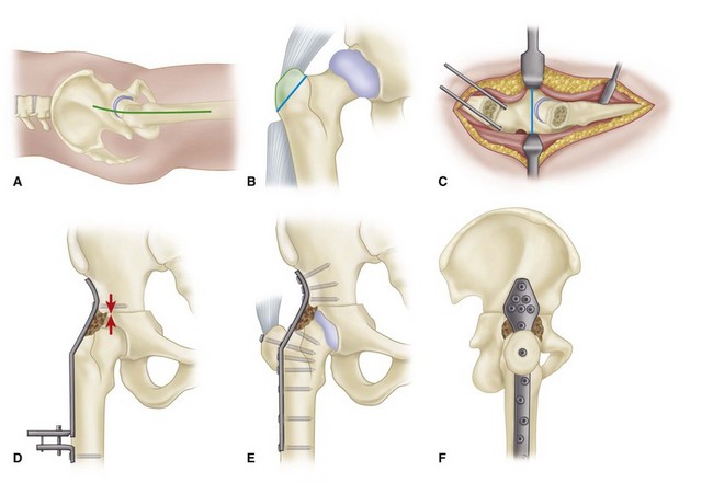
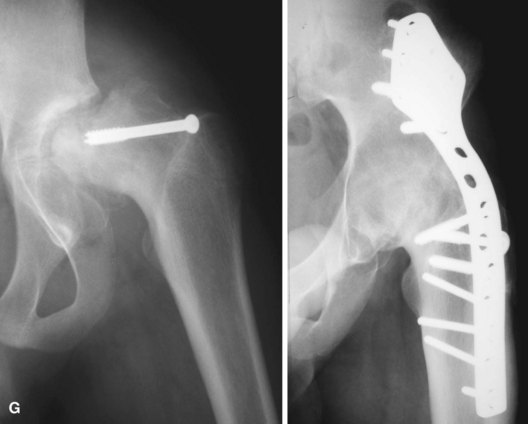

image No postoperative immobilization is applied.


Only gold members can continue reading. Log In or Register to continue

Stay updated, free articles. Join our Telegram channel

Jun 5, 2016 | Posted by in ORTHOPEDIC | Comments Off on Arthrodesis of the Hip

Full access? Get Clinical Tree

Get Clinical Tree app for offline access