35
Chronic Lacerations of Extensor Tendons
Angela A. Wang and Michelle Gerwin Carlson
History and Clinical Presentation
A 66-year-old right hand dominant man sustained a deep laceration to the dorsum of his right wrist when a plate glass mirror fell on it. He was evaluated at a local emergency room and was informed he would need surgery, but he wanted to be treated at another institution. Seventeen days later, he presented to the office with the complaint that he could not lift his fingers.
Physical Examination
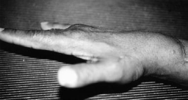
A well-healed 5-cm transverse laceration is noted on the dorsum of the right wrist just proximal to the edge of the extensor retinaculum. The patient lacked active extension of the index, long, and ring fingers at the metacarpophalangeal joints, as well as the thumb. In addition, he had a lack of extension of the wrist radially. The abductor pollicis longus, extensor pollicis brevis, and the small finger extensors were intact. Sensation in the distribution of the superficial radial nerve was intact (Fig. 35–1).
PEARLS
- Ultimate results are closely tied to postoperative care and rehabilitation. Careful attention to good splinting and controlled mobilization of the repaired tendon will yield better function when the tendon healing is complete.
PITFALLS
- The ruptured tendon must be repaired under the proper tension. Although tendon reconstruction under excessive tension is to be avoided, the surgeon must fix the tendon at the proper length to provide adequate power. It should also be noted that repairs usually loosen over time.
Diagnostic Studies
Radiographs of the right wrist were negative for bony pathology. No foreign bodies were present.
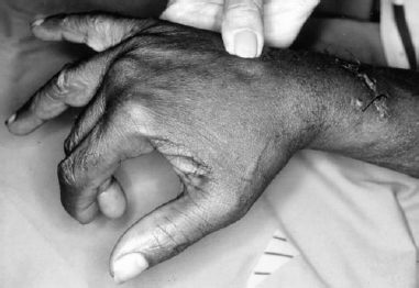
Figure 35–1. The patient under anesthesia preoperatively, with the wrist supported. Note the laceration on the dorsum of the wrist, and the lack of extension of the digits.
Differential Diagnosis
Extensor tendon laceration
Rheumatoid arthritis synovitis
Nerve injury
Diagnosis
Laceration of the Second, Third, and Fourth Dorsal Compartments of the Right Wrist, Late
Extensor tendon lacerations or ruptures that are treated in a delayed fashion may not be amenable to primary approximation and repair. Most tendon laceration and ruptures are best treated acutely; if this is not possible, the patient should be told that tendon reconstruction might be necessary. In cases of multiple tendon injuries, emphasis should be placed on recovering independent wrist and thumb extension, and mass extension of the fingers.
Postoperative care plays an important role in recovery. Early controlled mobilization (passive digital extension with wrist flexion and active digital flexion with wrist extension) and dynamic splinting have been shown to have positive effects on tendon healing and ultimate function.
Surgical Management
Four days later (3 weeks after the injury), the patient was brought to the operating room. Under regional anesthesia (an axillary block) with a tourniquet, an incision was made over the healed laceration and extended proximally and distally. The retinaculum was incised longitudinally over the fifth compartment, reflected back radially, and the dorsal compartments were identified and explored. The extensor carpi radialis brevis, extensor carpi radialis longus, extensor pollicis longus, extensor indicis proprius (EIP), and extensor digitorum communis (EDC) tendons were found to be lacerated. The extensor digitorum communis tendon had retracted proximally, and a large gap was present at the laceration site.
The extensor carpi radialis brevis and extensor carpi radialis longus tendons were directly repaired with a 4-strand repair using a 4–0 nonabsorbable, nonbraided suture. Similarly, the extensor pollicis longus was primarily repaired. Due to the chronic nature of the injury, however, direct approximation of the EIP and EDC tendons created significant tension and did not allow the digit to be flexed with the wrist in neutral. The decision was made to perform a fractional lengthening of the EIP and EDC tendons at the musculotendinous junction cutting the tendinious portion of the musculotendinous junction, after which the wrist could be brought into the neutral position and the fingers could be passively flexed. The wound was closed in a layered fashion over a Penrose drain. The patient was then placed in a volar splint with the wrist in 30 degrees of dorsiflexion and the fingers in 20 degrees of flexion at the metacarpalphalangeal joints.
Postoperative Management
On postoperative day 6, the patient was changed from his static splint to a dynamic outrigger extension splint, and began occupational therapy. The dynamic splint was discontinued at 3 weeks, whereupon the patient continued both active and passive range of motion exercises. He returned to work 9 weeks after the surgery, and by 3 months he had recovered full range of motion of the wrist and fingers, as well as good strength in the hand. (Figs. 35–2 and 35–3).