CHAPTER 40 Cervical Radiculopathy
Anterior Surgical Approach
Radiographic Evaluation
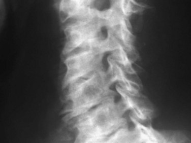
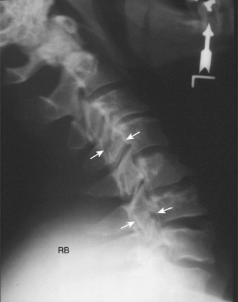
Radiographic evaluation usually begins with plain radiographs; lateral, anteroposterior, and oblique views are obtained. Global and segmental alignment can be easily assessed. Findings of degenerative disease include disc space narrowing, osteophytosis of the apophyseal joints, and spurring at the level the disc space. Degenerative spondylolisthesis of the cervical spine is common, and this usually involves facet arthropathy as well.1 Dynamic lateral radiographs can show segmental instability by assessing anteroposterior translation and angular motion. Foraminal stenosis may occur from uncovertebral arthrosis or degenerative subluxation. Typically, bony elements are visualized, but radiographs inherently are unable to assess neural elements. An indirect assessment of the neural foramina can be obtained with oblique views (Fig. 40–1). Although the soft tissue component of spinal cord compression cannot be visualized on lateral radiographs, the space available is easily assessed. Brain and Wilkinson2 reported an average space of 17 to 18 mm (range 14 to 23 mm).
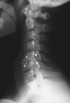
Several authors have reported on the use of lateral radiographs to assess cervical stenosis. A classic study by Edwards and LaRocca3 found the average sagittal diameter of asymptomatic individuals to be 17 mm. These authors reported that myelopathy was present in subjects with a diameter of less than 10 mm. A diameter of 10 to 13 mm was noted in patients without symptoms but who were likely to become symptomatic. Using radiographs, stenosis is present in about 5% of the population, and the rate increases with age to approximately 9% in patients older than 70 years.4 Torg ratio identifies stenosis and eliminates the variability and magnification associated with direct measurement of radiographs.5 A sagittal spinal canal diameter–to–vertebral body ratio of less than 0.8 is indicative of cervical spinal stenosis (Fig. 40–2). Several authors have questioned the reliability of Torg ratio.6–8
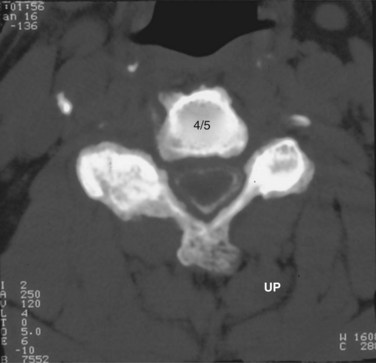
When more direct visualization of the neural elements is necessary, advanced imaging can be employed. Initially, spinal myelography was the test of choice. Water-soluble dye injected into the epidural space allows visualization of the neural elements by assessing flow patterns. Despite relatively high sensitivity, myelograms are nonspecific. Flow can be altered by soft disc herniations, hard disc (osteophyte formation), facet arthropathy, or various other pathologies. Computed tomography (CT) combined with myelography can significantly improve accuracy.9 Axial images and sagittal reconstructions improve the practitioner’s ability to determine the causes of compression. Altered filling of the nerve roots from bony compression can easily be assessed because of the bony detail produced with CT imaging (Fig. 40–3). CT myelography can be performed in patients who are not candidates for other procedures such as magnetic resonance imaging (MRI). Myelography has some disadvantages. A major concern is the invasiveness and morbidity associated with placement of the dye into the epidural space. Although CT myelography is more specific than radiographs, it is still very nonspecific for soft tissue causes of stenosis.
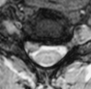
MRI offers several advantages. The neural elements and soft tissues are directly visualized, no radiation is required, and it is noninvasive. Meticulous inspection of the images can reveal soft disc herniations (Fig. 40–4) and sequestered fragments when they are present. Disruption of the posterior longitudinal ligament on sagittal MRI is indicative of a possibly sequestered fragment. Although MRI is best at visualizing soft tissue elements of the cervical spine, bony anatomy can be assessed. Additionally, MRI recognizes infiltrative soft tissue and vertebral body processes such as sarcoma or metastatic disease.
MRI scans offer excellent visualization of the lower brainstem and entire cervical spinal cord. The foramen magnum can be examined for Arnold-Chiari malformations. Basilar invagination or pannus formation from inflammatory arthritis can also be seen. Acute or chronic spinal cord compression may lead to signal change within the spinal cord, termed myelomalacia (Fig. 40–5). These changes may be important in predicting clinical outcomes after decompression.10,11
Although imaging has advanced greatly in recent years, a thorough history and physical examination still constitute the cornerstone of diagnosis and treatment. MRI and CT myelography must be correlated with the clinical evaluation of the patient. Boden and colleagues12 reported degenerative disc space changes in 25% of asymptomatic subjects younger than 40 years and almost 60% of subjects older than 60. A 10-year longitudinal study of healthy volunteers revealed that degenerative changes progressed, but symptoms developed in only 34% of subjects. Age was the only variable associated with the progression of spondylosis.13
Electrodiagnostic Studies
Electrodiagnostic studies are another diagnostic modality used to evaluate patients with radiculopathy. Peripheral nerve root entrapment can mimic cervical radiculopathy. Currently, electromyography (EMG) is the most useful modality. In some studies, 75% sensitivity has been reported.14–16 Because of the variable sensitivity of EMG, it is not an ideal screening tool. The diagnosis and location of pathology can be assessed with EMG, however. Compression causes pathophysiologic changes within the nerve root. These changes are shown on EMG as decreased motor unit potentials and fibrillation potentials. When attempting to distinguish between radiculopathy, peripheral neuropathy, and nerve root entrapment, neurophysiologic testing can be useful. When the diagnosis is clear from imaging and clinical examination, EMG is probably unnecessary. EMG and nerve conduction studies may be beneficial to distinguish specific nerve root compression when multilevel disease is present.
Surgical Indications
The indications for operative treatment of radiculopathy have not changed much over the years despite advances in operative techniques and devices. The most commonly accepted indications are (1) persistent or recurrent arm pain that is unresponsive to a trial of conservative treatment (3 months), (2) progressive neurologic deficit, (3) static neurologic deficit associated with significant radicular pain, and (4) confirmatory imaging studies consistent with the patient’s clinical findings. This chapter focuses on anterior approaches to surgical treatment. Smith and Robinson17 were the first to popularize the anterior approach.
Anterior Cervical Discectomy and Fusion: Surgical Technique
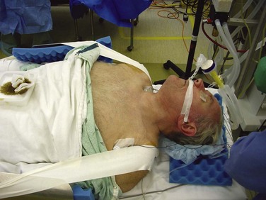
General endotracheal anesthesia provides the safest environment for anterior cervical discectomy and fusion (ACDF). The endotracheal tube should be taped on the side of the mouth opposite to the side on which the procedure is performed. Proper positioning is an essential part of this procedure. The authors typically place a small rolled towel longitudinally between the scapulae to allow the shoulders to fall back out of the way; this also positions the neck in mild hyperextension. Care must be taken in patients with myelopathy because iatrogenic worsening of myelopathy may occur with hyperextension. If symptoms are significantly worsened with hyperextension during the preoperative physical examination, this position should also be avoided. The arms are placed along the patient’s side and tucked. The shoulders can decrease visualization during exposure and during imaging. The authors use surgical tape to place longitudinal downward traction on the shoulders (Fig. 40–6).
More recently, it has been suggested that retractor displacement of the larynx against the endotracheal tube causes crush injury to the intralaryngeal segment of the recurrent laryngeal nerve, causing dysphonia. Apfelbaum and colleagues18 recommended deflating the endotracheal tube during the procedure to decrease pressure on the nerve and decrease the incidence of postoperative voice changes. Audu and colleagues19 performed a randomized, prospective, double-blind investigation of endotracheal tube manipulation. Postoperative examination with indirect laryngoscopy showed a 3.2% incidence of vocal cord paralysis. Endotracheal tube deflation did not reduce the incidence of vocal cord immobility. These authors concluded that tube manipulation has no effect on vocal cord injury.
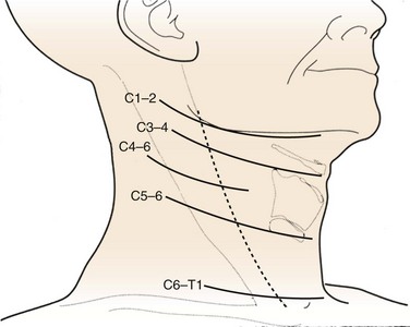
When a one-level or two-level discectomy is planned, a transverse incision is usually sufficient. These incisions tend to be more aesthetically pleasing. When an extensile approach is necessary, a longitudinal incision may be necessary. Patients with short, thick necks or who are undergoing revision surgery may benefit from longitudinal incisions. Fluoroscopy or superficial landmarks can be used to mark the appropriate skin level (Fig. 40–7). Typically, C3 is found at the level of the hyoid, C4-5 is found at the thyroid cartilage, and C6 is found at the cricoid cartilage. When exposing near the cervicothoracic junction (C7-T1), the inferior thyroid vessels and thoracic duct may have to be managed. A longitudinal incision may facilitate exposure at this level.
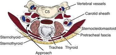
After the skin incision is complete, hemostasis can be obtained along the skin edges with electrocautery. The two edges of the skin can be elevated with either hand-held retractors or a small self-retaining retractor. The first muscle encountered is the platysma, which underlies the fascia. A curved hemostat is used to dissect bluntly underneath the platysma. Using electrocautery, the muscle is incised in line with the incision. Visualization is improved by opening the fascial planes underneath this layer; this also facilitates retractor placement to improve mobilization of the underlying structures. This is a crucial step in performing a safe dissection. The sternocleidomastoid should become visible. The dissection proceeds along the anteromedial aspect of the sternocleidomastoid. A blunt dissection should proceed between the carotid sheath and the trachea and esophagus. A finger should bluntly separate the medial border of the carotid sheath from the lateral border of the trachea and esophagus. Typically, the surgeon can easily discern the medial border of the tubular carotid. The pulsation of the carotid may not always be easily felt (Fig. 40–8).
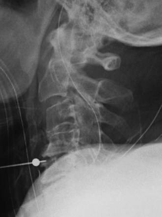
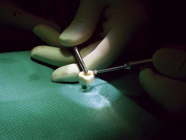
Correct identification of the proper level is imperative. Careful examination of radiographs may reveal anterior osteophytes. Palpation of these osteophytes on the anterior vertebral bodies can often lead to correct level identification. Lateral to C6 lies Chassaignac tubercle, which can be palpated and helps identify C5-6. Current patient safety recommendations by the North American Spine Society (NASS) recommend an intraoperative radiograph to verify the proper level. A standard spinal needle or specialized cervical marker should be placed in the intended operative level (Fig. 40–9). Identifying the level on a lateral radiograph may be difficult in the lower cervical spine or when the patient has either a short, thick neck or broad shoulders. The surgeon may elect to place a spinal needle at two levels, such that the superiormost needle can be visualized on the radiograph. Caution must be exercised if placement of two needles is required. A retrospective radiographic analysis of 87 consecutive patients by Nassr and colleagues20 showed a threefold increased risk of developing degenerative changes in disc levels that were previously marked with a needle for intraoperative level verification.
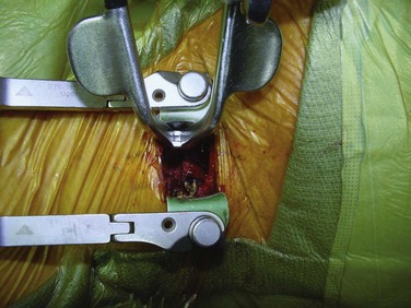
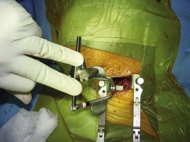
After the correct disc space is identified, the prevertebral fascia over the anterior vertebral body is divided; the longus colli muscles are then easily visualized. Dissection should begin in the midline and proceed laterally. The longus muscles are elevated bilaterally with a combination of electrocautery and mild blunt dissection. The exposure is held open with self-retaining retractors that are placed under the longus colli muscles (Fig. 40–10). The retractor can be secured by an assistant or with an arm secured to the operating table.
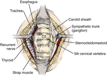
FIGURE 40–10 After elevation of longus colli muscles bilaterally, blunt retractors can be placed for remainder of procedure.
Visualization of the posterior half of the disc can be impaired by overhanging anterior osteophytes. These can be removed with a bur or rongeur to improve visualization (Fig. 40–11). It is imperative that the surgeon note the angle of the disc space on the intraoperative localization film, and care should be taken to perform the disc removal along the angle of the endplates to avoid inadvertent penetration of the vertebral bodies. The uncinate process, posterior longitudinal ligament, and bony endplates should be visible at the end of the discectomy. Inspection of the posterior longitudinal ligament is necessary to determine if a sequestered fragment is present.
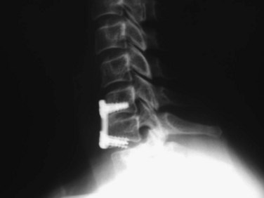
The significance of posterior osteophyte removal is unknown. When they are causing obvious compression on the spinal cord, osteophytes should be removed (Fig. 40–12). After the neural elements have been decompressed and the endplates have been prepared, the anterior to posterior depth of the disc should be measured with a calibrated nerve hook or a small wire. Generally, the disc space is 10 to 15 mm wide, 12 to 17 mm deep, and 5 to 10 mm long. Regardless of the type used, the intervertebral graft should be several millimeters less than the actual depth to allow recession and avoid spinal cord compression.
The endplates should be punctured down to bleeding bone to facilitate bony union; various ways exist to accomplish this. Some surgeons prefer to use a bur to remove the endplates partially, whereas others use a small, angled curet to create holes in the adjacent sides. Regardless of the method, excessive endplate removal must be avoided. Endplate collapse and graft subsidence can occur when the structural integrity is weakened too much. When using tricortical iliac crest or patella, the graft should be shaped in a lordotic fashion with the anterior height being 1 to 2 mm taller than the posterior height (Fig. 40–13). This shape helps to avoid posterior retropulsion of the graft and subsequent cord compression and restores lordosis. The graft should be placed with the disc space under distraction to facilitate insertion. Longitudinal traction by the anesthesiologist is as effective as various intervertebral distractors. A tamp and mallet are used to position the graft (Fig. 40–14).
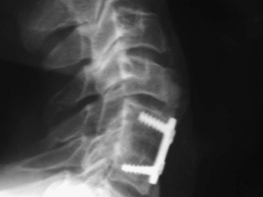
If an anterior cervical plate is not a planned part of the procedure, the bone graft is usually countersunk 1 to 2 mm in relation to the anterior margin of the adjacent vertebral bodies to decrease the chance of graft extrusion and esophageal compression (Fig. 40–15). When adding a plate, only minimal graft countersinking is necessary. A final lateral intraoperative radiograph is strongly suggested. Correct graft position can be verified along with proper plate position. The authors typically place a drain just anterior to the vertebral body. The fascia overlying the platysma is closed with simple absorbable sutures, and the skin is closed with a running absorbable subcuticular suture.
Consensus on postoperative management of patients after ACDF is lacking. Initially, most surgeons required patients to wear a rigid cervical collar for 6 to 8 weeks. Anterior instrumentation has eliminated many graft-related complications. Some surgeons have abandoned or significantly limited their use of postoperative immobilization because of the safety and effectiveness of current instrumentation. More recent studies have shown that a postoperative cervical brace does not improve the fusion rate or the clinical outcomes of patients undergoing single-level anterior cervical fusion with or without plating.21,22
When adequate internal fixation is obtained, a soft collar can be used for a short time in the initial postoperative period. Modern perioperative care and techniques allow patient mobilization on the day of surgery and discharge on postoperative day 1. Some surgeons have shown similar clinical outcomes and safety when performing ACDF in the outpatient setting.23–25 At 1 to 2 weeks, follow-up anteroposterior and lateral radiographs are taken. Lateral radiographs show a sufficient incorporation of the bone graft such that patients are allowed to begin range of motion exercises and strengthening exercises by 6 weeks. Within 3 months of the surgical procedure, most patients can expect return to full activity.
Anterior Cervical Plating
Anterior cervical plating was first introduced in the 1960s, when Bohler25a modified lower extremity hardware. Initially, he used the plates in trauma cases to restore stability. Exponential advancements have been made in the manufacture of anterior cervical instrumentation, biocompatibility, and ease of implantation. Currently, the use of an anterior cervical plate has been advocated to increase postoperative stability, decrease the incidence of graft extrusion, decrease the incidence of graft collapse, improve fusion rate, and obviate the need for postoperative halo brace placement after extensive multilevel corpectomies.
Biomechanical testing of bicortical anterior cervical instrumentation after single-level discectomy has shown a marked reduction in motion compared with the intact specimen.26 Additional testing by Grubb and colleagues27 showed that the Cervical Spine Locking Plate System (Synthes, Paoli, PA) using unicortical screws is biomechanically equivalent and in some cases more stable than the bicortical screw Caspar system. These results were noted after fixation of an experimental severe compression flexion injury in the cervical spine. The study by Spivak and colleagues28 showed that locking screws significantly increase rigidity of the hardware systems initially and after cyclic loading. Based on a combination of biomechanical and clinical studies at this time, most surgeons recommend the use of unicortical plating systems.
The clinical effectiveness of anterior cervical plate fixation must be judged not only by the successful fusion rate, but also by an improved clinical outcome. Over the past decade, several investigators have reported prospective and retrospective studies, looking at the two groups of patients fused with and without anterior cervical plates.29 Connolly and colleagues29 reported on 43 patients treated for cervical spondylosis with ACDF using autogenous iliac crest bone graft. The authors noted that there was no difference in the overall fusion rate or clinical results between these two groups of patients (plate vs. no plate).
Samartzis and colleagues30 reviewed 69 consecutive patients who had undergone single-level ACDF with and without rigid instrumentation. The nonplated group consisted of 38 patients, and the plated group consisted of 31 patients. All patients had tricortical iliac crest autograft as the interbody. The plated group wore hard collars for 3 to 4 weeks, and the nonplated group wore hard collars for 6 to 8 weeks. Fusion rates of 100% and 90% were achieved in the nonplated and plated groups. These rates were not significantly different. Mean blood loss was significantly more in the plated group. Clinical success was assessed using Odom criteria at an average of 21 months. Excellent and good outcomes was reported in 91.3% and 92.1% in the nonplated and plated groups. Three nonfused patients reported good clinical outcome. Of patients, 24 were smokers, and 23 had work-related injuries. Smokers achieved a fusion rate of 95.8%. Clinical outcome and fusion rate were similar between the groups in regard to smoking status and work-related injuries.
Current systems have incorporated the use of dynamic locking. Theoretically, these systems decrease stress shielding and allow load sharing to occur between the graft and the anterior column. The decrease in graft loading reduces the risk of plate or screw failure by recreating the natural load sharing of the anterior column.31–33 Some designs involve “dynamic screws,” and others involve “dynamic plates.”
Several more recent studies have shown that no significant clinical differences exist between static and dynamic plates.34–36 Pitzen and colleagues34 performed a prospective, controlled, randomized, multicenter study of 132 patients undergoing an anterior cervical discectomy and autograft fusion with either a dynamic plate or a rigid plate. They monitored patients for implant complications, segmental mobility, fusion, loss of lordosis, and clinical outcomes (visual analog scale [VAS] and Oswestry Disability Index [ODI]). No implant complications were seen in the dynamic group, whereas four implant complications occurred in the static group. There was no difference in fusion rate or clinical outcomes at 2 years. The loss of segmental lordosis was significantly greater in the dynamic group. By the criteria of Pitzen and colleagues,34 the dynamic group achieved fusion at a faster rate. These authors recommended using dynamic plates because of fewer implant-related revisions and a faster time to fusion.
Nunley and colleagues35 conducted a prospective, randomized study of static versus dynamic plates in single-level and multilevel ACDF. All patients were treated for radiculopathy. Multilevel procedures were performed in 38 of the 66 patients. When considering all study subjects, clinical outcome was equivalent between the dynamic and static groups. A clinically significant reduction in VAS and neck disability index (NDI) occurred in 73% of patients. A solid fusion was observed in 85% of patients. Clinical success predicted radiographic fusion, but fusion did not predict VAS and NDI reduction. Dynamic plates showed significantly better clinical outcomes in multilevel fusions than static plates. Nunley and colleagues35 concluded that plate design has no effect on clinical outcome in single-level constructs. Patients undergoing multilevel fusions experience better clinical outcomes with dynamic plating, however. The loss of lordosis may be related more to graft settling than to plate design, but this has not compromised outcomes.37
McLaughlin and colleagues38 detailed a cost analysis of 64 patients who underwent two-level ACDF for cervical radiculopathy. This study compared the cost of rigid internal fixation for patients with two-level ACDF with the cost in a similar population of patients who had an identical procedure without instrumentation. No difference was noted in the clinical outcome between the two groups, with both groups obtaining a 92% excellent or good result using Odom criteria. The investigators noted that patients who underwent rigid plating returned to light activities, driving, and unrestricted work sooner than noninstrumented patients. Although the overall cost was increased by approximately $3500 for the patients in the instrumentation group, the investigators believed that the increased cost was justified, owing to the significant clinical advantage to the patients and insurance disability providers, secondary to earlier mobilization and return to work.
Caspar and colleagues39 and Geisler and colleagues40 described one of the largest studies examining the results of plate fixation after ACDF. These two articles, describing the same group of patients, included 365 patients who underwent a one-level or two-level cervical arthrodesis performed with and without anterior cervical plate stabilization. The intended goal of this study was to determine the incidence of a second surgical intervention and whether the use of an anterior cervical plate decreased the need for subsequent surgical procedures. Although patients in these studies were not randomly assigned, and there was a mix of autograft and allograft procedures and multilevel fusions, the investigators reported that in the entire group 22 patients required repeat surgical intervention, with 20 of these patients initially in the noninstrumented group.
The addition of an anterior cervical plate using unicortical screw fixation after ACDF rarely leads to additional intraoperative complications. Plate fixation usually follows graft placement with care being taken to countersink the graft only minimally below the adjacent endplates. Typically, the exposure required for an ACDF is sufficient for placement of the cervical plate. Anterior osteophytes at the superior and inferior endplates need to be removed to provide a flat surface for plate placement. Placement of the cervical plate on top of anterior osteophytes may lead to prominence of the hardware with subsequent difficulty with postoperative swallowing. Unicortical screw placement can usually proceed without the use of fluoroscopy, with screw placement directed medially and away from the involved disc space (Fig. 40–16). Care should be taken to select a plate of the appropriate size so that the adjacent disc spaces are not violated by the screw (Fig. 40–17).
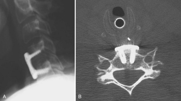
The authors recommend placing the plate at least 5 mm away from the adjacent disc space to decrease the risk of ossification (Fig. 40–18).41 Proper unicortical screw placement minimizes the risk of spinal cord injury. Intraoperative vertebral artery injury is extremely rare and occurs only with extreme screw misplacement. If sufficient surgical exposure has been obtained for the discectomy portion of the procedure, placement of the anterior cervical plate usually adds only 15 to 20 minutes to the entire operative time.
The use of an anterior cervical plate after a one-level ACDF is now common. Although the successful fusion rate may be higher with the use of instrumentation, clinical outcome has not yet been shown to be significantly improved with the addition of this hardware. As the complexity of cervical reconstructive surgical procedures increases, the use of cervical plates seems to be justified, owing to the higher fusion rate with instrumentation at two-level and three-level disease.42–44 Wang and colleagues44 compared the clinical and radiographic outcomes of 60 patients after two level ACDF with and without plating. The pseudarthrosis rate was significantly different at 25% for the nonplated group and 0% in the plated group. Age, gender, levels of surgery, tobacco use, and prior surgery had no correlation with fusion. A significant amount of segmental kyphosis and graft collapse was noted in the nonplated group. The complication rates were similar, however. Regardless of instrumentation, 87% of fused patients showed good to excellent results. Of patients with a pseudarthrosis, 58% reported fair results, and only 28% reported good to excellent results.
Results
In 1962, Robinson and colleagues45 were the first to publish the results of a large series of patients treated with ACDF. The series consisted of 55 patients who underwent anterior discectomy and fusion at 107 disc spaces. These authors hypothesized that preservation of the subchondral bone of the endplates was necessary to provide a bearing surface for the graft. (Emery and colleagues46 challenged this hypothesis and noted a pseudarthrosis rate of only 4.4% when partially removing the subchondral bone of the endplate with a bur.) Robinson and colleagues45 reported 46% excellent, 27% good, 22% fair, and 6% poor results. Patients with single-level fusions showed better outcomes than patients with multilevel fusions. More than 94% of patients with single-level fusions had an excellent or good result compared with 50% of patients undergoing three or more levels of fusion. Four patients continued to report symptoms that were believed to be associated with nonunion, and there were nine total pseudarthroses. Most importantly, only one additional surgery was necessary in a patient with a solid fusion.
Yue and colleagues47 performed a long-term retrospective review of 71 patients after ACDF using allograft and plating. The average follow-up was 7 years (range 5 to 11 years). Single-level and multilevel patients were included. More than 90% were primary procedures. Symptoms included neck pain (94%), radiculopathy (91%), and weakness or gait problems (79%). Greater than 95% of patients reported continued significant improvement in neck pain and radiculopathy at final follow-up. Clinical improvement was not related to fusion, smoking, number of levels, collapse, or subsidence. The pseudarthrosis rate was 9%. Smoking had no effect on fusion rate. Previous surgery increased the risk of pseudarthrosis, however. No patient underwent revision for pseudarthrosis. Adjacent segment degeneration occurred in 73% of patients. Only 16% required additional surgery, with half exhibiting degenerative changes before the index procedure. Global lordosis was increased with surgery and maintained at final follow-up.
Despite a large volume of literature reporting high fusion rates with single-level ACDF, multilevel procedures present a challenge for the spine surgeon. Fusion and clinical outcomes decrease with increasing levels of surgery. Emery and colleagues48 retrospectively reviewed 16 patients who underwent a modified Robinson ACDF at three operative levels. All patients had tricortical autogenous iliac graft placed at three levels with no internal fixation. Only 9 (56%) of the 16 patients went on to achieve solid arthrodesis at all levels. Of the seven patients with pseudarthrosis, two had severe pain requiring revision, two had moderate pain, and three had no pain. Of the nine patients with a solid fusion, three continued to have mild pain, and six had no pain. Emery and colleagues48 concluded that a three-level modified Robinson ACDF results in an unacceptably high rate of pseudarthrosis and recommended that these patients undergo additional or alternative measures to achieve a successful arthrodesis consistently.
Wright and Eisenstein 49 compared fusion rates in single-level and two-level anterior discectomy and fusion without plating. In the 54 single-level procedures, pseudarthrosis occurred in 11% compared with 28% in the two-level group. Postoperative neck pain was less likely in patients with a solid fusion.
In an effort to improve the successful fusion and patient outcome after multilevel ACDF, Bolesta and colleagues50 reported a prospective study of 15 patients who underwent a modified Smith-Robinson ACDF at three and four operative levels, stabilized with a unicortical anterior plate. All patients had autogenous iliac crest graft placed and had a minimum of 24 months of follow-up. Despite the addition of the anterior cervical plate, solid arthrodesis was achieved at all levels in only 7 (47%) of the 15 patients after the initial surgical procedure. Of the eight patients with pseudarthrosis, three had sufficient pain to necessitate revision surgery, one had pain without further surgery, and four had no pain. Of the seven patients with solid fusion, three had persistent pain, and four had none. Bolesta and colleagues50 also concluded that a three-level and four-level modified Robinson ACDF results in an unacceptably high rate of pseudarthrosis. These authors believed that the addition of an anterior cervical plate alone does not improve the arthrodesis rate.
Wang and colleagues44,51 also studied multilevel procedures. They reviewed 60 patients after two-level ACDF.44 Half of the patients received plate fixation, and half did not. The difference in fusion was significant. No pseudarthrosis occurred in the plated group, whereas seven pseudarthroses occurred in the nonplated group. The complication rate was similar between the groups. Pseudarthrosis was not related to demographics or smoking status. During this same period, Wang and colleagues51 reviewed three-level procedures as well. Of 59 patients followed for 3.2 years, pseudarthrosis occurred in 18% of plated patients versus 37% of nonplated patients. This difference was not significant, however, owing to the larger number of plated patients. Fused patients showed better clinical outcomes regardless of plating. Cauthen and colleagues52 reviewed 146 multilevel noninstrumented cases at an average of 5 years and found a 75% fusion rate. Fusion rates are lower but still acceptable for multilevel procedures when plating is employed.
The clinical effect of pseudarthrosis on patient outcome is controversial. Patients who have a successful fusion theoretically have improved cervical alignment, continued foraminal distraction, and prevention of collapse into kyphosis. The pseudarthrosis rate after single-level surgery ranges from 0% to 20%53,54 with the rates approaching 50% for patients undergoing multilevel surgery.55,56
Phillips and colleagues57 attempted to determine the natural history, risk factors, and treatment outcomes in a large population with documented pseudarthrosis after ACDF. They reported 48 patients with radiographically proven pseudarthrosis at an average of 5 years after the initial surgical procedure. Of the patients, 67% were symptomatic at latest follow-up or at the time of a required second surgical intervention. Several patients had a symptom-free period of up to 2 years after ACDF before redeveloping cervical symptoms after a minor traumatic episode. Analysis of the results revealed that patients who had surgery at a younger age had an increased likelihood of pseudarthrosis becoming symptomatic. Of 16 patients, 14 had successful repair of pseudarthrosis via an anterior approach, whereas 6 patients underwent posterior cervical fusion, with all going on to successful fusion. In patients in whom fusion was achieved with a second cervical operation, the results were excellent in 19 and good in 1. Phillips and colleagues57 concluded that the surgical repair of pseudarthrosis with either an anterior or a posterior approach results in a higher likelihood of a successful clinical outcome.
The etiology of pseudarthrosis after anterior cervical surgery is multifactorial. Various authors have cited multilevel fusions, tobacco use, and revision cervical surgery as relative risk factors for the development of pseudarthrosis. Multiple studies of lumbar spine fusions have shown that cigarette smoking creates a biologically challenging environment for a successful fusion. Hilibrand and colleagues58 compared the long-term radiographic and clinical results of smokers and nonsmokers who had undergone arthrodesis with autogenous bone graft after multilevel anterior cervical decompression for the treatment of cervical radiculopathy or myelopathy or both. After either corpectomy or multiple discectomies and interbody grafting, 190 patients were followed clinically and radiographically for at least 2 years. A subset of 40 of these patients were smokers who had undergone a cervical fusion, and only 20 had a solid fusion at all levels, whereas 69 of the 91 nonsmokers had a solid fusion at all levels (P < .02). This difference was more significant among patients who had a two-level interbody grafting procedure. Hilibrand and colleagues58 concluded that smoking had a significant negative impact on healing and clinical recovery after multilevel ACDF with autogenous interbody graft for radiculopathy or myelopathy.
A prospective study by Luszczyk and colleagues59 analyzed the fusion rates of smoking and nonsmoking patients who underwent single-level ACDF with allograft and a rigid locked anterior cervical plate. The study consisted of patients from the control groups of four separate studies evaluating cervical disc arthroplasty; 156 smokers and 417 nonsmokers were reviewed. Follow-up was at least 24 months in all patients. When analyzing all the patients, the fusion rate was 91.4%. The fusion rates were similar for the two subgroups at 91% and 91.6% for smokers and nonsmokers. These authors concluded that smoking status had no effect on the fusion status in single-level ACDF when using allograft and a locked plate.
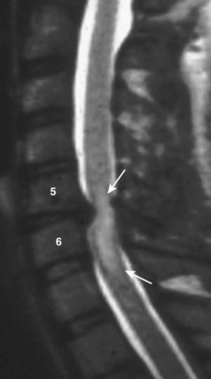
Long-term follow-up studies of patients treated with ACDF have shown that approximately 10% of patients undergo reoperation at an adjacent level, owing to progressive spondylolysis or a new disc herniation (Fig. 40–19).60–63 This adjacent segment disease has been theorized to be secondary to the increased biomechanical stresses on an unfused level adjacent to a solid fusion. Whether the biomechanical forces alone are sufficient to cause new disease or this is just a progression of the natural history of cervical spondylosis in a patient with significant degenerative changes remains to be determined. Treatment of this adjacent level disease is theoretically more difficult, however, because achievement of a solid fusion in these patients is hindered by the long lever arm of the prior fusion acting across the new operative level.
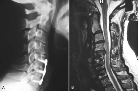
FIGURE 40–19 A and B, Lateral radiographs and sagittal MRI showing disc herniation adjacent to two-level fusion.
Related posts:

Stay updated, free articles. Join our Telegram channel

Full access? Get Clinical Tree


