Those not associated with a systemic illness
Photosensitive drug eruptions
Photoallergic contact dermatitis
Polymorphous light eruption and its variants
Solar urticaria
Those that can be associated with a systemic illness
Cutaneous LE
Cutaneous dermatomyositis
Porphyria/pseudoporphyria
Some photosensitive disorders can display skin changes in areas not directly exposed to natural (sunlight) or artificial forms of ultraviolet light (e.g., cutaneous dermatomyositis, cutaneous LE, eczematous or lichenoid photosensitive drug eruptions) as well as in photoexposed areas. Typically, the rash starts in the areas of skin directly exposed to ultraviolet light and then spreads to contiguous nonexposed areas. Other photosensitive disorders characteristically produce skin involvement limited to areas directly exposed to ultraviolet light (e.g., polymorphous light eruption, solar urticaria, photoallergic contact dermatitis).
The patient denied using any over-the-counter topical products likely to contain contact-sensitizing chemicals (neomycin, bacitracin, diphenhydramine). Therefore, it is likely that the observed skin changes are the expression of the primary disease process rather than secondary changes produced by allergic contact dermatitis.
Clinical Context. The patient’s Past Medical History includes mild hypertension over the past 5 years currently controlled with medical therapy. For the past 10 years, the patient had been under medical care for gastroesophageal reflux disease. The patient has a 20-year history of hypothyroidism. Review of Systems – The patient admitted to mild joint pains predominantly in her wrists and fingers over the past 3 months. She had also recently noticed the onset of malaise and easy fatigue upon exertion. Social History – The patient has smoked one-half pack of cigarettes daily for the past 30 years. Family History – The patient’s mother had a history of alopecia areata and her younger sister developed vitiligo as a youth. Current Medications – Hydrochlorothiazide, lisinopril, omeprazole, and levothyroxine. Medication Allergies – None known.
Personal analysis of clinical context findings. Medical disorders such as hypertension and acid reflux disease are often treated with drugs that have the potential to cause photosensitive adverse skin reactions. Several of the medications that this patient is taking for her other medical problems fall into this category (e.g., hydrochlorothiazide, lisinopril, and omeprazole). In addition, these same drug classes have been reported to be capable of triggering drug-induced SCLE.
Early-onset hypothyroidism often results from autoimmune thyroid disease such as Hashimoto’s thyroiditis. Individuals have had one end-organ autoimmune disease like autoimmune thyroiditis that is linked to the 8.1 ancestral HLA haplotype are at risk for developing other diseases that are linked to this haplotype (e.g., vitiligo, alopecia areata, SCLE, Sjögren’s syndrome, type 1 diabetes mellitus, Addison’s disease, pernicious anemia) [2].
The patient’s recent onset of mild arthralgia, malaise, and easy fatigue would suggest the presence of a photosensitive skin disorder that is associated with systemic manifestations such as a cutaneous LE or cutaneous dermatomyositis rather than photosensitive skin disorders that are typically not accompanied by systemic inflammation (see Table 1.1).
If the patient proves to have a form of cutaneous LE, her history of cigarette smoking could result in a suboptimal clinical response to aminoquinoline antimalarial therapy [3].
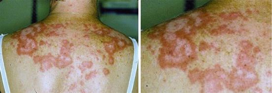
Physical Examination. The patient was asked to disrobe and put on a hospital gown. The patient had papulosquamous skin lesions of varying size and shape distributed symmetrically on the lateral aspects of her neck, the V area of her upper chest, her shoulders, her upper back, the extensor surfaces of her distal arms, the extensor surface of her forearms, and the dorsal aspects of her hands. The smaller lesions were papulosquamous (i.e., red and scaly) papules and small plaques. However, the larger lesions were ring-shaped (i.e., annular) lesions with erythema and scale at the active edges and the absence of such changes centrally. The inactive centers of the lesions displayed a white-gray hue (i.e., leukoderma, meaning a decrease in or absence of melanin pigment) compared to the noninvolved perilesional skin (Fig. 1.1). In some areas, the annular lesions merged producing a polycyclic arrangement of lesions (Fig. 1.1).


Fig. 1.1
Annular SCLE lesions. The right panel is an enlargement of the left upper quadrant of the clinical shown in the left panel. Note the light color of the skin within the inactive central parts of the annular lesions. Also note the polycyclic arrays resulting from the merging together of the larger annular lesions on the posterior aspects of the patient’s shoulders
There was no obvious dermal scarring associated with any of these skin changes. In addition, there was no periungual erythema on her fingers nor any grossly visible periungual microvascular abnormalities. Bedside capillaroscopy with a dermatoscope failed to reveal any significant periungual microvascular abnormalities. In addition, there were no grossly visible cuticular abnormalities including hypertrophy or disarray. There was no tenderness, erythema, or swelling of the small joints of her hands and fingers. The ocular and oral mucosal membranes were not involved.
Personal analysis of physical examination findings. In a patient having a chronic rash of unknown etiology, it is important to have the patient disrobe and put on an examination gown so that a complete skin evaluation can be performed. Attention should be paid to pertinent negative findings as well as pertinent positive findings during the exam. For example, our patient indicated that her rash did not occur below her waist. However, subtle skin changes of disorders that can produce changes below the waist such as cutaneous dermatomyositis can be missed if the patient is not examined completely (e.g., patchy violaceous erythema over the lateral hips [holster sign], subtle violaceous erythema over the knees and medial malleoli). Most forms of cutaneous LE do not produce changes below the waist.
In addition, inflammatory skin changes on one part of the body can at times be secondary to a focus of skin inflammation on another part of the body. As an example, patients with inflammatory skin changes on their feet resulting from dermatophyte fungal infection can develop aseptic eczematous skin changes over their upper extremities and back as a result of the dermatophytid reaction (a fungus-triggered autoeczematization reaction) [4]. One can misinterpret the cause of the rash on the arms and back in this setting if one does not examine the feet to recognize the appropriate etiologic association.
Stay updated, free articles. Join our Telegram channel

Full access? Get Clinical Tree


