Fig. 18.1
Reactive arthritis – erosions on tongue (Reprinted from Wu IB, Schwartz RA. Reiter’s syndrome: The classic triad and more. J Am Acad Dermatol 2008;59:113–121. With permission from Elsevier)
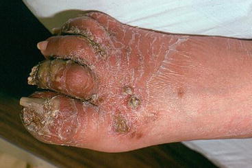
Fig. 18.2
Nail changes include nail dystrophy, subungual debris, and periungual pustules. (Reprinted from Wu IB, Schwartz RA. Reiter’s syndrome: The classic triad and more. J Am Acad Dermatol 2008;59:113–121. With permission from Elsevier)
KD accompanies ReA and is one of its cardinal clinical manifestations, although the vast majority of patients with ReA never demonstrate cutaneous features and are never seen by a dermatologist. It is estimated that KD occurs in about 10 % of ReA patients.
Differential Diagnosis
The differential diagnosis for keratoderma blennorrhagica in the genital area includes Bowen’s disease, candidiasis, Paget’s disease, contact dermatitis, squamous cell carcinoma, erosive lichen planus, lichen sclerosus, fixed drug eruption, and, of course, psoriasis.
The differential diagnosis for lesions found on the palms and soles includes psoriasis as well as hyperkeratosis of the palms/soles, pustular eruption of the palms/soles, scabies, and dermatophytosis. Oral lesions of ReA may resemble aphthous ulcers, geographic tongue, lichen planus, candidiasis, and autoimmune bullous diseases.
Biopsy
KD lesions show spongiform macropustules in the upper epidermis, intraepidermal microabscesses, marked papillomatosis, and acanthosis. Older lesions show cornified and thickened layers and acanthosis. The histologic findings in KD are indistinguishable from psoriasis (as are the clinical features). Subungual hyperkeratosis, found in KD, may also be indistinguishable from psoriasis.
Histopathologic findings of the early cutaneous lesions are essentially the same as in psoriasis. Early lesions of keratoderma blennorrhagicum feature a spongiform pustule in the upper dermis. Later lesions of keratoderma blennorrhagicum usually do not contain spongiform pustules but reveal the nonspecific findings of acanthosis, hyperkeratosis, and parakeratosis.
Diagnostic Flow Chart
The diagnosis of KD and other dermatologic lesions in ReA is made by obtaining a complete medical history and thoroughly examining the patient. The constellation of symptoms that confirm the clinical diagnosis of ReA may not occur contemporaneously and the clinician must take a very careful history. No laboratory test confirms a diagnosis of ReA. HLA-B27 testing is neither diagnostic nor confirmatory, although its presence does portend a possibly more aggressive form of the disease. The erythrocyte sedimentation rate (ESR) is often elevated as is the C-reactive protein (CRP). Leukocytosis and an anemia of chronic inflammation may be seen. The rheumatoid factor (RF) and antinuclear antibody (ANA) are both absent. If early in the disease course, one might be able to culture Chlamydia from the urine or an enteric pathogen from the stool. HIV and syphilis testing should always be considered. Imaging studies of affected joints, at least in the early phase of symptoms, usually add little useful clinical information.
Related posts:

Stay updated, free articles. Join our Telegram channel

Full access? Get Clinical Tree


