Fig. 43.1
Sharply demarcated erythematous patch on the calf (Courtesy of Orhan Aral, Istanbul University, Division of Rheumatology)
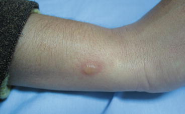
Fig. 43.2
Bullae on an erythematous base on the forearm (Courtesy of Ayşe Akman-Karakas, Akdeniz University, Department of Dermatology)
Differential Diagnosis
Erythematous patches resembling FMF lesions may be caused by other dermatoses. Erysipelas and cellulitis are common streptococcal infections of skin causing erythematous warm patches and plaques accompanied with fever and chills [6]. In contrast to ELE these lesions do not subside in a few days spontaneously but respond to systemic antibiotic treatment. Sweet’s syndrome presents with fever, systemic symptoms, and long-lasting well-demarcated erythematous nodules and plaques with some common histopathological features with ELE. Lesions of Sweet’s syndrome are often multiple and elevated, and histopathologically the dermal edema is prominent. These lesions are not confined to lower extremities, and treatment with systemic corticosteroids is preferred [10]. Other hereditary recurrent fever syndromes, for example, HIDS (hyperimmunoglobulinemia D syndrome) and TRAPS, with various skin manifestations are included in the differential diagnosis of FMF [11]. HIDS begins usually under 1 year of age and the presence of high serum level of IgD is diagnostic. Various types of skin manifestations including maculopapular and papular eruptions are seen nearly in all of the patients [1]. Histopathologically a mild vasculitis may be observed. Skin manifestations are common in TRAPS and may be generalized in contrast to FMF. Large erythematous migratory patches and swollen plaques with indistinct borders are accompanied by painful myalgia. The rash shows perivascular infiltration of lymphocytes and monocytes [1, 11].
Biopsy
The histopathological features of ELE and nonspecific skin lesions are not diagnostic for FMF [11]. Histopathological examination of ELE shows slight edema of the superficial dermis, a sparse mixed perivascular infiltration composed of lymphocytes, neutrophils, histiocytes, and nuclear dust. Vasculitis is not seen. Direct immunofluorescence examination of these patches reveals deposits of C3 in the wall of superficial vessels. IgM and fibrinogen can be detected in some patients. The nonspecific bullous lesions show a subepidermal cleavage without any specific immunoglobulin deposition in the direct immunofluorescence examination [6].
See Also
Henoch-Schonlein purpura, neonatal onset autoinflammatory diseases (TRAPS), Behçet’s disease.
Explanation of Algorithm
Major Criteria*
1.
Peritonitis (generalized)
2.
Pleuritis or pericarditis
3.


Monoarthritis (hip, knee, ankle)
Related posts:

Stay updated, free articles. Join our Telegram channel
Full access? Get Clinical Tree
