Fig. 23.1
Dusky cyanosis from vasoconstriction in Raynaud’s phenomenon (Reprinted with from Wigley FM, Wung PK. Painful Digital Ulcers in a Scleroderma Patient with Raynaud’s Phenomenon. In: Silver RM, Denton CP. (eds). Case Studies in Systemic Sclerosis. Springer-Verlag. London, UK; 2011: 95–105. With permission from Springer Science + Business Media)
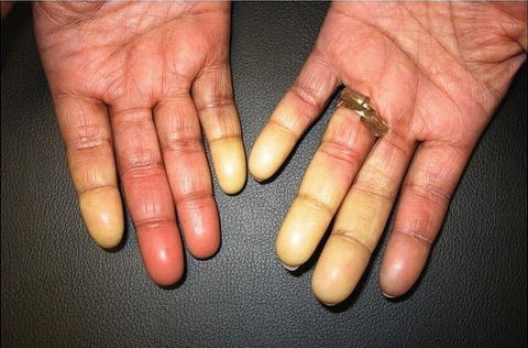
Fig. 23.2
Sharply demarcated pallor on several digits in the acute ischemic phase of Raynaud’s phenomenon (Courtesy of Noëlle S. Sherber, Fredrick M. Wigley)
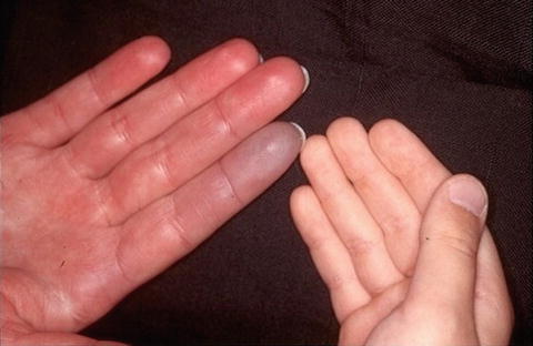
Fig. 23.3
Hyperemia in the blood flow recovery phase of Raynaud’s phenomenon, with coexistent cyanosis of an adjacent digit, as compared to an unaffected hand (Courtesy of Noëlle S. Sherber, Fredrick M. Wigley)
During the ischemic phase, patients often experience numbness and paresthesias as a result of digital vessel vasospasm. True pain is a symptom of deprived tissue nutrition and is a warning symptom of potential impending tissue injury. Ischemic ulcerations can result from tissue injury (Fig. 23.4). An uncomplicated ischemic event is typically over in about 15 min.
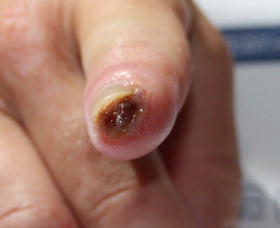
Fig. 23.4
Ischemic digital ulceration resulting from tissue injury in Raynaud’s phenomenon (Reprinted with from Wigley FM, Wung PK. Painful Digital Ulcers in a Scleroderma Patient with Raynaud’s Phenomenon. In: Silver RM, Denton CP. (eds). Case Studies in Systemic Sclerosis. Springer-Verlag. London, UK; 2011: 95–105. With permission from Springer Science + Business Media)
Differential Diagnosis
Raynaud’s phenomenon (RP) may be an exaggerated normal response of the cutaneous thermoregulatory vessels and digital arteries to cold, stress, or trauma (primary Raynaud’s phenomenon), or it can be associated with an underlying disease state, such as scleroderma (secondary Raynaud’s phenomenon). Primary Raynaud’s phenomenon is characterized by the absence of an underlying systemic disorder and is observed in approximately 3–5 % of individuals in the United States [1–3].
A frequent cause of secondary Raynaud’s phenomenon is an underlying systemic rheumatic disease. The highest prevalence of RP among patients with a rheumatic disease is observed in systemic sclerosis (scleroderma) or in mixed connective tissue disease (MCTD), approaching 90 % or greater in most studies [4–7]. It is estimated that approximately 50 % of patients with undifferentiated connective tissue disease (UCTD) [8, 9], 21–44 % of patients with SLE [10, 11], up to 17 % of patients who have rheumatoid arthritis [12], and approximately 10 % of patients who have polymyositis demonstrate Raynaud’s phenomenon as well.
Secondary Raynaud’s can also be induced by trauma (hand-arm vibration syndrome), mechanical obstruction (thoracic outlet syndrome), pathologic serum proteins (cryoglobulinemia, cryofibrinogenemia), neurogenic stimuli (carpal tunnel syndrome), and exogenous vasoconstrictors (chemotherapeutic drugs).
Biopsy
There is no pathognomonic histology of Raynaud’s, but, in disease states such as scleroderma, intimal hyperplasia of the digital vessels is characteristic.
References
1.
Gelber AC, Wigley FM, Stallings RY, et al. Symptoms of Raynaud’s phenomenon in an inner-city African-American community: prevalence and self-reported cardiovascular comorbidity. J Clin Epidemiol. 1999;52:441–6.PubMedCrossRef
Related posts:

Stay updated, free articles. Join our Telegram channel

Full access? Get Clinical Tree


