Fig. 33.1
(a)“Peau d’orange” feature. The superficial dermis may be wrinkled while the deep dermis and fascias are fibrosed. (b) Indentation along the course of the superficial veins (groove sign)
Biological tests show peripheral eosinophilia and increased inflammatory markers (elevated sedimentation rate and C-reactive protein), at least during the acute phase of the disease. Muscular enzymes may be mildly increased reflecting the underlying muscular involvement. Autoimmune anaemia and hypergammaglobulinemia (sometimes monoclonal gammopathy) are variably present. Immunological markers of autoimmunity such as antinuclear antibodies or rheumatoid factors have not been reported in EF with consistency.
Magnetic resonance imaging of the involved limbs is able to detect fascial thickening and precise the extension of the lesions especially in the muscles (Fig. 33.2). Abnormal signal intensity and contrast enhancement are suggestive of an active inflammation. These findings are useful to make the diagnosis, to guide the location for biopsy and to monitor the response to therapy [8]. High-resolution ultrasonography appreciates the extension of the lesions in the dermis. Although these preceding examinations are very useful for managing patients, attempt for confirmation of the diagnosis has to include the examination of a full-thickness skin to muscle biopsy.
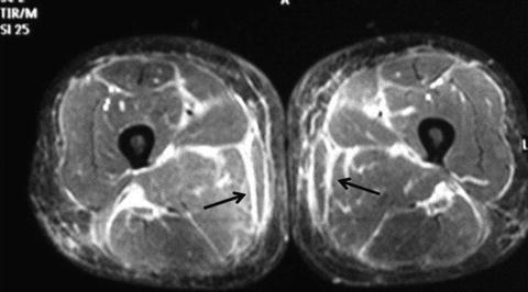
Fig. 33.2
Thigh muscle MRI of a patient with eosinophilic fasciitis. Axial fat-suppressed, T2-weighted fast spin-echo MR image shows markedly increased signal intensity within the superficial and deep fascial layers and mildly increased T2 signal intensity within the superficial muscle fibres adjacent to the fascia (arrows)
Differential Diagnosis
Systemic sclerosis, localised scleroderma, inflammatory myositis, hypereosinophilic syndrome, graft versus host disease, toxic oil syndrome or eosinophilia–myalgia syndrome are considered as differential diagnoses of EF. Lyme disease, vasculitis and cutaneous T-cell lymphoma might also be considered. The most important parameters to be considered for differential diagnosis are listed in Table 33.1.
Table 33.1
Differential diagnoses of eosinophilic fasciitis
Systemic sclerosis | Presence of Raynaud phenomenon, capillaroscopic abnormalities, antinuclear antibodies, visceral involvements; fibrosis mainly localised in the dermis |
Morphea or localised scleroderma | All the areas of the body may be involved. Usually, fibrosis is mainly localised in the dermis; however, in some cases, fibrosis may involve the fascias and the muscles as in EF, especially in the linear forms affecting the limbs. So, EF is frequently considered as a deep variant subset of morphea |
Inflammatory myositis and dermatomyositis (DM) | Proximal muscular weakness, general feeling of discomfort, DM skin manifestations, high levels of muscle enzymes, MRI examination and inflammation predominantly localised in the muscles |
Hypereosinophilic syndrome | Sustained absolute eosinophil count greater than >1,500/μl, manifestations of organ involvement (heart, central nervous system, skin, respiratory tract…) |
Eosinophilic–myalgia syndrome and toxic oil fibrosis | Epidemic disorders resulting from ingestion of toxic contaminants such as aniline-denaturate rapeseed oil and l-tryptophan; sudden onset with more severe manifestations: myalgias, oedema and multi-organ involvement (mainly the lung and central nervous system) leading to a high mortality rate |
Sclerodermoid graft versus host disease (GVH) | Onset after allogeneic stem cell transplantation, association with lichen sclerosus and morpheaform lesions mainly on the trunk with sometimes EF-like lesions on the limbs; association with liver abnormalities and gut involvement; initial fibrosis in the superficial dermis [9]
Related posts:Stay updated, free articles. Join our Telegram channel
Full access? Get Clinical Tree
 Get Clinical Tree app for offline access

|
