Fig. 9.1
Typical erythema nodosum of the lower legs (a) and an atypical macular form (b)
However, erythema nodosum may be atypical, being either widespread small, macular mimicking cutaneous vasculitis or the neutrophilic dermatoses such as Sweet’s syndrome (Fig. 9.1b) or diffuse periarticular and mimicking arthritis (Fig. 9.2). Involvement of the upper extremities alone is very rare for erythema nodosum. Known associations and triggers of erythema nodosum include intercurrent viral illnesses; streptococcal, mycobacterial, and yersinia infection; oral contraceptives; antibiotics; SLE and other connective tissue diseases; Crohn’s disease; and primary biliary cirrhosis. In many cases of erythema nodosum, an associated trigger or disease will not be identified, up to 20 % in one series [3].
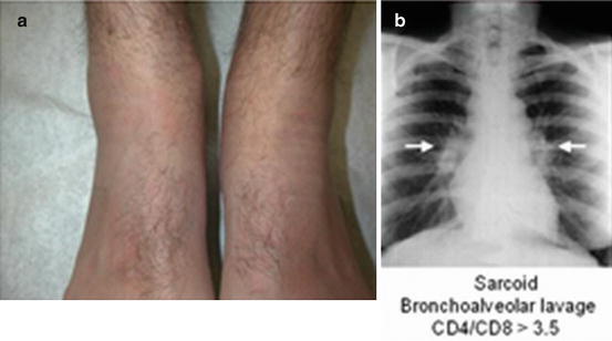
Fig. 9.2
Diffuse periarticular erythema nodosum on the right ankle – a chest X-ray showed large, bilateral adenopathy (arrows) consistent with Loeffler’s syndrome (sarcoidosis)
Another nonvasculitic, mostly lobular panniculitis is the rheumatoid nodule. Typically occurring on pressure areas such as the elbows (Fig. 9.3a) with a distinct histology (Fig. 9.4), they may occur anywhere and mimic gouty tophi.
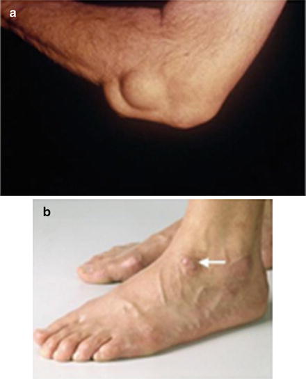
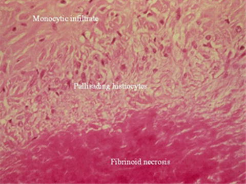
Fig. 9.3
Rheumatoid nodule on elbow (a), histologically identical to granuloma annulare on dorsum of foot in another patient (b)
Fig. 9.4
Histology of a rheumatoid nodule
Two other lesions have a similar histology to the rheumatoid nodule: granuloma annulare [4] and necrobiosis lipoidica diabeticorum. The former may be referred to a rheumatologist following biopsy of isolated nodular skin lesions, usually on the dorsum of the foot in a young patient following an intercurrent viral respiratory infection (Fig. 9.3b). Awareness of this self-limiting benign condition will avoid unnecessary biopsies and other investigations.
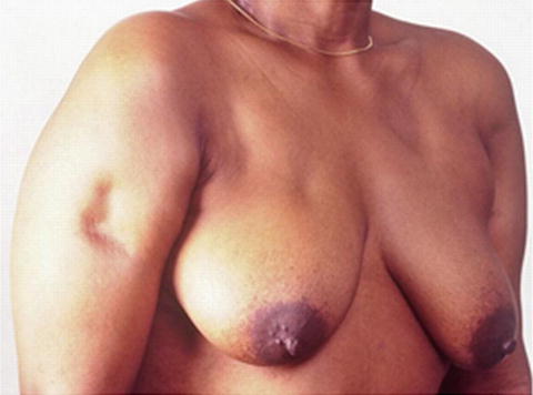
An unusual form of panniculitis seen in <2 % of SLE patients is lupus profundus [5]. Occurring on the trunk and proximal extremities, this nonvasculitic, painless lesion is mostly lobular and evolves to atrophy (Fig. 9.5).