Fig. 51.1
The “Bassett ligament,” also named “meniscoid lesion,” is a type of anterolateral fibrous impingement. It is caused by an accessory fascicle of AITFL that has sometimes a vertical direction for a more distal insertion on the fibula
In the “classic” AAI, anterior spurs were supposed to be traction osteophytes of the anterior capsule due to repetitive plantar flexion movements of the ankle during kicking [2]. The anterior joint capsule attaches actually more proximally to the tibia and more distally to the talus compared to the location of the spurs, and arthroscopy of the ankle allows to clearly inspect and to remove them without opening the capsule. It has been demonstrated through an experimental study [13] that in football players, the impaction-related microtrauma due to the repetitive ball impacts on the anterior aspect of the ankle can lead to the formation of hyperostosis in the distal tibia and in the talus through the cascade of events described above for the pathogenesis of anteromedial impingement (fibrocartilage and secondary formation of calcification and hyperostosis).
51.1.3 Clinical and Diagnostic Examination
The symptoms of an AAI are usually a chronic anterior ankle pain and some kind of swelling after sports activity with a history of recurrent ankle sprains. Many times a slightly limited dorsal flexion can be detected. Anterolateral, anterior, or anteromedial impingement can be distinguished according to the site of the pain during activities and on palpation during physical examination.
The radiographic examination is essential for the diagnosis of the bony impingement. In the classic anterior impingement (the “footballer’s ankle”), a lateral view of the ankle can detect the presence of hyperostosis of the anterior profile of the distal tibia and/or of the anterior border of the talar dome: the spurs can pinch the synovia during dorsiflexion and provoke pain and swelling (Fig. 51.2).
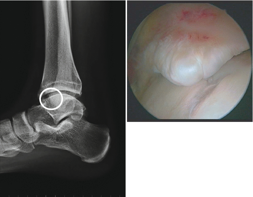
Fig. 51.2
“Classical” anterior ankle impingement. Presence of an anterior tibial spur seen on a radiographic lateral view and during an anterior arthroscopy (“the footballer’s ankle”)
In anteromedial impingement, the presence of hyperostosis on the anterior aspect of the medial malleolus and on the medial border of the talus can be undetected on standard radiographs because of the prominence of the anterior border of the distal tibia and of the dorsal profile of the neck of the talus. Van Dijk et al. [14] described an anteromedial radiographic oblique view (described in Fig. 51.3) that can detect the anteromedial hyperostosis and allow the correct diagnosis.
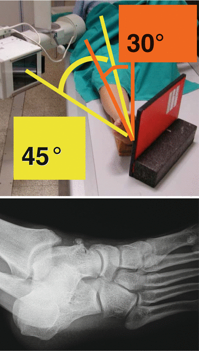
Fig. 51.3
The radiographic oblique view to detect an anteromedial bony impingement. The leg is positioned in 30° of external rotation; the radiographic beam is 45° oblique on the longitudinal axis of the leg, perpendicular to the radiographic film, centered on the ankle
Anterolateral impingement cannot be detected with radiographs as it is a soft tissue impingement. The diagnosis is done on clinical data: in a recent paper [15], the efficacy of conventional MRI in detecting soft tissue abnormalities in anterolateral gutter remains controversial, with a wide range of sensitivities (39–100 %) and specificities (50–100 %).
51.1.4 Treatment Strategy
Pain in anterior ankle impingement seems to be due to the entrapment of the synovial tissue during the movements of the ankle, mainly in dorsal flexion. The presence of hyperostosis can reduce the anterior articular space and may facilitate this pathologic mechanism. Arthroscopy is theoretically the most useful tool to remove bony spurs and the synovial pathological tissue having fewer possibilities to create hypertrophic scars that might be painful again.
Anterior arthroscopy is not a trivial kind of surgery: accuracy is essential in positioning the patient, in performing the sites of entry portals, and in exchanging the arthroscope and instruments for the treatment of different types of impingement. Without dealing with the general technique of anterior ankle arthroscopy, we would like to emphasize some specific tips to consider in the treatment of an AAI.
Position of the patient: the patient must be supine, with the foot on the border of the table in order to be able to perform passively dorsal or plantar flexion during surgery. The foot must be exactly vertical on the plane of the table, and the operated leg must be slightly elevated compared to the other one to allow comfortable maneuvers of the instruments.
Sites of the portals: anteromedial portal is performed near the medial border of the tibialis tendon with the ankle in full dorsal flexion. A more medial site may create difficulties in viewing the lateral malleolus in case of lateral ossicles or peroneal hyperostosis for the convexity of the talar dome. The lateral portal is identified by the scope with a needle, on the lateral edge of the peroneus tertius muscle, avoiding the superficial peroneal nerve by dissecting the subcutaneous layer with a mosquito clamp. Also this portal must be performed as “central” as possible to allow a comfortable position of the instruments to work on the medial malleolus if it were required. If an anterolateral soft tissue impingement is suspected, it is recommended to locate this portal exactly at the joint level; to address a pathology of the medial malleolus, it is more useful to locate the anterolateral portal slightly more proximal in order to view more easily the malleolus [16].
Scope and instruments: after a routine exploration of the joint and the confirmation of the type of pathology, the portals must be used according to the pathology to treat. In anteromedial impingement, medial ossicles, hyperostosis of medial malleolus and of the neck of the talus, the medial portal is the working portal while the arthroscope is shifted in the anterolateral portal. In lateral pathology (anterolateral soft tissue impingement, removal of peroneal ossicles or osteophytes), instruments must be kept in the lateral portal. To remove an anterior bony impingement (tibial and/or talar), scope and instruments can be interchanged to address the spurs conveniently. It is not always clear how much bone must be resected to treat the bony impingement: tibial hyperostosis must be identified proximally to fully define the anatomic shape of the anterior tibia. In this way the proximal limit of the bony spur can be clearly addressed with a burr, performing the debridement in a proximal-distal direction. For the talar spurs, the distal base of implant of the spur must be identified to make a distal-proximal resection.
51.2 Posterior Ankle Impingement
51.2.1 Etiology and Classification
Posterior ankle impingement (PAI) is a painful syndrome in the posterior aspect of the ankle, and it is a mechanical conflict that arises in forced plantar flexion.
The talus extends posteriorly with a bony process that can cross laterally the midline in case of hypertrophy: it is called the posterolateral process, and in about 7 % of the adult population, it appears as a distinct accessory bone for a failed union of this ossification center [17]. This center is normally separated from the ossification center of the body of the talus until 8–10 years of life. In adults this distinct ossicle, if present, is called os trigonum, and it is usually linked to the talus through a fibrous synchondrosis. Medially to the posterolateral process, there is another smaller bony prominence of the talus, the medial tubercle: between these two bony prominences, there is a sulcus that is transformed in an osteofibrotic channel by a fibrous retinaculum that takes insertion on the tip of the two tubercles. The flexor hallucis longus (FHL), the most posterior of the posteromedial tendons, slides in this channel. At this level the channel acts as a pulley where the FHL changes its direction of sliding, from vertical to nearly horizontal: this is a point of weakness of the tendon, and pathologies that can involve the posterolateral talar process (or the os trigonum, if present) can also involve the tendon itself and cause a FHL tendinitis. The posterolateral process also gives insertion to the posterior talofibular ligament (PTFL), the strong and most posterior ligament of the lateral ligamentous complex of the ankle. This ligament is lax in plantar flexion and taut in maximal dorsal flexion, and for this reason, it can detach the process (or the os trigonum) from the talus during forced and sudden movements.
51.2.2 Injury Mechanism
PAI can be caused by trauma or overuse, and it has been typically associated with ballet dancers for the frequent “en pointe” or “demi-pointe” position assumed by these athletes. However, any sport that requires or any trauma that provokes forced plantar flexion or more rarely forced dorsal flexion can determine this syndrome. Running (especially downhill running), soccer, basketball, volleyball, and jumping (high, long, and triple jump) are the sports more frequently involved in this kind of pathology. Rarely the detachment of the posterior bony process can be caused by a sudden traction of the PTFL during a trauma in forced dorsal flexion. More frequently a forced plantar flexion of the ankle, sometimes in association with supination or pronation of the hindfoot, provokes a crush of the os trigonum or of the posterolateral process between the posteroinferior edge of the tibia and the calcaneus. The mechanism of this crush has been compared to a nut (the os trigonum) in a nutcracker (tibia and calcaneus). This kind of trauma can be acute and can determine the fracture of the posterior talar process or the sudden mobilization of the os trigonum. Other times the trauma can be repetitive during a long period of time and can cause an overuse syndrome. This different etiology is not academic, but it is of practical interest because of better prognosis of PAI in overuse syndromes compared to traumatic cases [18]. The presence of the os trigonum or of a hypertrophic posterolateral process is neither sufficient nor essential to determine this syndrome. If the bony process or the ossicle is present, they are not sufficient to determine the pain syndrome: sometimes the pain is caused only by the traumatic event or by efforts beyond the anatomical limit. In other patients the painful syndrome is caused by a soft tissue impingement: a thickened joint capsule, a rupture of the PTFL, and a bump of hypertrophic tissue may cause a PAI syndrome without bony prominence.
Related posts:

Stay updated, free articles. Join our Telegram channel

Full access? Get Clinical Tree


