Anatomical ACL Reconstruction with BPTB Autograft via Rectangular Tunnels A single-bundle technique based on the concept of double-bundle reconstruction
Konsei Shino
Shigeto Nakagawa
Tatsuo Mae
INTRODUCTION
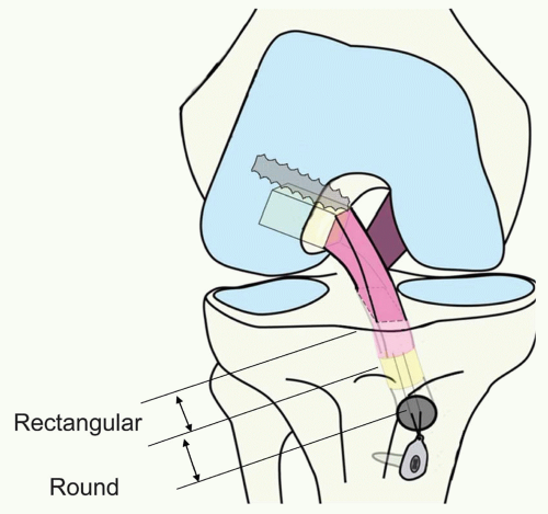
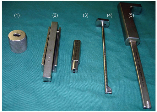
The autogenous bone-patellar tendon-bone (BTB) of 10-mm width has been one of the most frequently used ACL grafts to reconstruct as a single bundle via round tunnels (6,8). As the rectangular shape of its cross section is close to that of the natural ACL, it could fairly mimic the natural fiber arrangement inside the ACL if it is properly placed in the tunnels created in the right attachment areas (Fig. 27.1) (2,4). In order not only to mimic the natural fiber arrangement inside the ACL, but to maximize the graft-tunnel contact area in the femur as well as in the tibia, this rectangular tunnel technique was developed (Fig. 27.2). Consequently, this procedure follows the concept of the double-bundle reconstruction (11). The instruments were developed in cooperation with Smith & Nephew Endoscopy (SNE), Andover, Massachusetts (Fig. 27.3).
SURGICAL TECHNIQUE
Positioning and Skin Incision
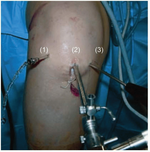
Using a leg holder, the distal thigh is kept horizontal to obtain a consistent view of the intercondylar notch, regardless of knee flexion angle. A skin incision of 3 in is made along the medial border of the patellar tendon, and then curved medially to the entry point of the tibial tunnel, located just above the pes anserinus (Fig. 27.4).
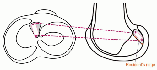 FIGURE 27.1 Placement and orientation of the BTB graft in the right knee. Points A and B in the femoral attachment correspond A′ and B′ in the tibial attachment, respectively. |
Graft Harvesting and Preparation
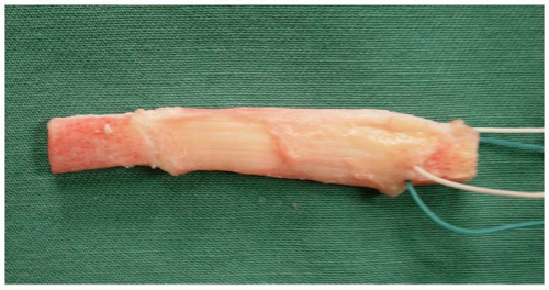
A 10-mm wide BTB graft is harvested from the medial half of the patellar tendon with 15-mm long bone plugs on both ends. This offers the graft’s tendinous portion with uneven length. The medial longer side is assigned to the anteromedial bundle, while the central shorter one is for the posterolateral bundle.
The bone plug from the tibia is shaped with the bone-plug shaper (SNE#72200447) into a rectangular parallelepiped socket of 5 mm thick × 10 mm wide × 15 mm long to snugly pass the graft sizing template (SNE#6901101) and used for the femoral socket. The patellar bone block is left as a triangular pillar for the tibial tunnel (Fig. 27.5).
Arthroscopic Portals
In addition to routine anterolateral and anteromedial portals, the far anteromedial portal, 2 to 2.5 cm posterior to the anteromedial portal and just above the medial meniscus, is routinely created (9


Stay updated, free articles. Join our Telegram channel

Full access? Get Clinical Tree


