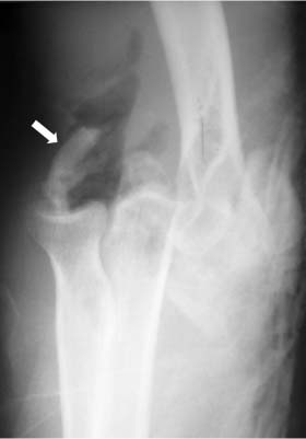
14 Acute Fracture-Dislocations about the Elbow Elbow dislocation is the second most common type of dislocation encountered in the adult upper extremity and constitutes 10 to 25% of all injuries to the elbow.1 When an isolated elbow dislocation occurs without an associated fracture (simple dislocation), closed reduction of the elbow with short-term immobilization and early range of motion (ROM; within 2 weeks) can provide a stable joint with good functional outcome.2–4 However, an elbow dislocation with concomitant intraarticular fracture of the radial head, olecranon, or coronoid process is termed a complex dislocation; its treatment requires strict adherence to a systematic stepwise treatment approach to obtain reliable patient satisfaction and functional results.5,6 Acute elbow fracture-dislocations are a challenge to treat because they are prone to acute recurrent instability, chronic instability, stiffness, posttraumatic arthrosis, and pain.7,8 Operative procedures for reconstruction of elbow fracture-dislocations are technically demanding because of the complex anatomy, the interplay between soft tissue and osseous injuries, and the proximity of crucial neurovascular structures. In this chapter, we will discuss the diagnosis, standard surgical approach, and rehabilitation of acute fracture-dislocations about the elbow. The elbow is an inherently stable joint. However, the stability of the elbow becomes compromised and more difficult to control as the soft tissue injury becomes more extensive or with associated fractures.9 As we have gained more experience treating elbow fracture-dislocations, it has become evident that careful attention must be paid to both the bony and soft tissue disruption associated with the injury. In 1987, Broberg and Morrey10 reported radiographic arthrosis in 22 of 24 patients with a fracture-dislocation of the elbow treated without repair or replacement of the radial head at an average of 10 years postoperatively. In 1989, Josefsson et al11 reported the long-term (3 to 34 years) outcome in 23 patients who had sustained an elbow dislocation with a displaced radial head fracture. They reported a high prevalence of osteoarthrosis in 12 of 19 patients who had been treated with radial head excision following elbow fracture-dislocation. In addition, they noted redislocation in 4 patients, all of whom had an untreated fracture of the coronoid process. In 1989, Regan and Morrey12 reported on the Mayo Clinic experience in 35 patients who had a fracture of the coronoid process of the ulna. In their series, treatment of type II fractures resulted in worse outcomes with an associated radial head fracture or elbow instability, and 4 of the 5 patients with a type III fracture had a poor result secondary to stiffness, pain, and recurrent instability. In 2002, Ring et al13 reviewed 11 patients with posterior dislocation and associated radial head and coronoid fractures, the so-called terrible triad of the elbow. Nine patients had the radial head treated by internal fixation (5 patients) or excision (4 patients), with reattachment of the lateral collateral ligament complex (LCLC) in only 3 patients. All coronoid fractures were less than 50% and were not fixed. Five elbows redislocated in the splint postoperatively including all four with radial head excision. The result of treatment was rated as unsatisfactory for 7 of the 11 patients. Together these studies highlight (1) the difficulties in the management of these complex injuries, (2) the reduction of the coronoid fragment is critical to restore elbow stability, and (3) resection of the radial head is contraindicated when the elbow is unstable. It is well recognized that prolonged immobilization following an episode of acute elbow instability is associated with poor outcome.4 The dilemma in management of complex elbow fracture-dislocations is that, without appropriate surgical reconstruction, instability rapidly ensues with attempted motion, but prolonged cast immobilization leads to unacceptable stiffness. In addition, immobilization in a cast does not ensure concentric reduction of the elbow.13 In previous studies, standard reconstruction protocols based on restoring both osseous and soft tissue structures were not used to optimize elbow stability, leading to high rates of prolonged cast immobilization and resultant stiffness and arthrosis.11,13,14 An improved understanding of the mechanism of elbow instability, the primary and secondary constraints providing stability, the patterns of soft tissue disruption, and improved techniques for surgical repair have led to the development of a surgical strategy for these injuries. The elbow is a complex hinge joint that relies on a combination of bony articulations and soft tissue constraints to optimize stability and mobility.15 The ulnohumeral articulation is the essential factor for osseous stability and mobility in the flexion-extension plane. It has been shown that the osseous articulation provides 30% resistance to valgus stress and 75% resistance to varus stress with the elbow at 90 degrees of flexion.15 The olecranon blocks the anterior translation of the ulna with respect to the distal humerus while an intact coronoid process provides an anterior buttress resisting posterior subluxation of the proximal ulna in extension beyond 30 degrees or greater.16 It has been shown that 50% of the coronoid is required to maintain stability against a direct posterior force in an otherwise intact elbow.17 The medial facet of the coronoid is especially crucial to stability in varus stress.18 The importance of the radial head as a secondary stabilizer to valgus stress and posterior translation is well recognized; however, the radial head also indirectly contributes to varus stability by creating tension in the lateral ligament complex.19 Schneeberger et al20 have recently evaluated the concurrent role that the radial head and the coronoid process have as posterolateral rotatory stabilizers of the elbow and the stabilizing effect of radial head replacement and coronoid reconstruction. The posterolateral rotatory displacement of the ulna was measured after application of a valgus and supinating torque in seven cadaveric elbows. Resection of the radial head with an intact LCLC showed a significant increase in rotatory instability compared with intact elbows. The clinical correlation of this finding was recently reported by Hall and McKee,21 who described posterolateral rotatory instability (PLRI) in 7 patients following radial head resection. Additional removal of 30% of the height of the coronoid fully destabilized the elbows, always resulting in ulnohumeral dislocation despite intact ligaments. The implantation of a rigid radial head without reconstruction of the coronoid restored stability to the elbow. However, the elbows with a defect of 50 or 70% of the coronoid, loss of the radial head, and intact ligaments could not be stabilized by radial head replacement alone, but additional coronoid reconstruction restored stability. This study highlights the importance of the radial head for elbow stability and further illustrates the necessity for coronoid reconstruction in more complex injuries. The treatment of elbow fracture-dislocations has traditionally focused on management of the bony injuries. However, it has become apparent that equal attention should be paid to the dynamic and static soft tissue stabilizers. Soft tissue constraints about the elbow are responsible for as much as 40 and 50% of the resistance to valgus stress and varus stress in the extended position, respectively.22 McKee et al23 have defined the patterns of soft tissue disruption in 61 patients (62 elbows) requiring operative repair for a dislocation (10 cases) or fracture-dislocation (52 cases) of the elbow. In the 52 fracture-dislocations, the associated fractures were coronoid (n = 39), radial head (n = 36), proximal ulna (n = 14), and distal humerus (n = 6). Disruption of the LCLC was seen in all 62 elbows in one of six patterns: proximal avulsion (52%), bony avulsion of lateral condyle (8%) (Fig. 14–1), midsubstance rupture (29%), ulnar detachment (5%), ulnar bony avulsion (5%), or combined (2%). Rupture of the common extensor origin was seen in 66% of the patients. Medial side injuries were confirmed in 80% of cases by direct visualization, intraoperative stress testing, or radiographs. The exact role that the dynamic stabilizers, such as the common extensors, play in providing stability to the elbow has not been clearly defined; however, active ROM of the elbow is inherently more stable than passive ROM.24 The anterior band of the ulnar collateral ligament acts as the major stabilizer to valgus stress. The major stabilizer to varus or rotatory stress is the LCLC, including the lateral ulnar collateral ligament.25,26 Figure 14–1 Acute disruption of the lateral collateral ligament complex (LCLC) occurs in one of six patterns. Although proximal soft tissue avulsion is the most common, bony avulsion (white arrow) can also occur as illustrated in this anteroposterior radiograph of a posterolateral elbow fracture-dislocation. Repair of the LCLC, posterolateral capsule, and common extensor origin is a critical step in the operative technique to obtain and maintain a stable congruent joint. Posterior dislocations of the elbow are commonly caused by a fall on the outstretched hand or wrist with either a hyperextension or posterolateral rotatory mechanism. In the hyperextension mechanism, an anterior force is generated that levers the ulna out of the trochlea as force is transmitted from the fall to the extended elbow. As the joint progressively hyperextends, the anterior capsule and collateral ligaments are placed under increasing tension and eventually fail. O’Driscoll et al have described a combination of valgus stress and forearm supination with axial compression through the lateral column of the elbow as a mechanism for elbow dislocation. This posterolateral dislocation causes a sequential soft tissue and possibly bony disruption from lateral to medial and has been classified into three stages. In stage 1, the LCLC is disrupted resulting in PLRI; stage 2 involves further soft tissue disruption of the anterior and posterior capsule resulting in incomplete posterolateral dislocation; and stage 3 involves further soft tissue disruption and instability subdivided into three degrees (3A, anterior band medial collateral ligament [MCL] intact; 3B, anterior band of MCL disrupted; 3C, entire distal humerus stripped of all soft tissue).26,27 The more uncommon anterior dislocation may be caused by impact on the posterior forearm in a slightly flexed position. Most acute elbow dislocations are posterior and involve both the radius and ulna. The distinction between posterior, posterolateral, and posteromedial is sometimes difficult to determine and seldom influences treatment. The other positions of dislocation – anterior, medial, lateral, and divergent – are rare and require individualized care.22 No single universally accepted classification scheme exists for elbow fracture-dislocations. However, the type of injury can be determined based on the injury acuity, direction of dislocation, and extent of ligamentous disruption and associated fractures. Radial head fractures are classified based on the modified Mason’s classification: type I fractures represent nondisplaced or minimally displaced fractures (less than 2 mm) of the radial head or neck; type II fractures are displaced fractures of the radial head or neck but fixable; and type III fractures are displaced radial head or neck fractures, but unreconstructable. Type IV injuries are defined as a fracture of the radial head with dislocation of the ulnohumeral joint.28,29 Failure to restore the contact of the radial head with the capitellum may compromise not only immediate elbow stability, but also the ability of the LCL and MCL complexes to heal with proper physiologic tension.9,29 Excision of the radial head without replacement can lead to progressive valgus instability, deformity, PLRI, and arthrosis in the setting of concomitant MCL injury. Therefore, the current recommendation is for reconstruction or replacement of the radial head in this setting.28,30,31 Associated proximal ulna fractures can involve the coronoid process and/or the olecranon process. The integrity of the coronoid process of the ulna plays a substantial, if not essential, role in elbow stability.17 Regan and Morrey12 classified fractures of the coronoid process into three types, depending on the extent of involvement. Type I fractures are a small fleck of bone sheared from the coronoid during subluxation or dislocation; type II fractures involve up to 50% of the coronoid process; and type III fractures involve more than 50% of the coronoid. Recently, Sanchez-Sotelo and colleagues18 have added an additional fracture pattern (type IV) to describe the sagittal plane fracture of the coronoid involving the attachment of the anterior bundle of the MCL. This medial oblique compression fracture occurs with an axial load and probable varus force. The depressed anteromedial fragment causes a pathognomonic double subchondral density on the lateral radiograph, which has been termed the double crescent sign. It has been suggested that 50% of the coronoid can be fractured before instability develops in the intact elbow situation; however, in the setting of an elbow dislocation, even type I coronoid fractures are important for stability. It has become apparent that even these small fragments provide important stability through their attachment to the anterior capsule.32 The subcutaneous position of the ulna makes it vulnerable to direct trauma that can result in complex fractures of the proximal ulna. In cases of severe force to the elbow, a fracture dislocation can occur with posterior displacement of the olecranon fragment and displacement of the distal ulnar fragment together with the head of the radius anterior to the humerus. An associated coronoid fracture is not infrequent and typically is triangular and involves 50 to 100% of the coronoid process. It is important to restore a stable trochlear notch for effective treatment of transolecranon fracture-dislocations of the elbow.33 In complex proximal ulna fractures with comminution, the tension band technique will collapse the fragments together leading to a narrowed olecranon articulation that does not track properly. The optimal fixation for these fractures is offered with contoured, limited-contract dynamic compression (LCDC) plate fixation, with or without bone graft depending on the size of the comminuted region.34 The advantages of using the LCDC plate for fixation are several-fold.35,36
Anatomy
Treatment
Etiology and Mechanism of Injury
Classification
Associated Radial Head Fractures
Associated Proximal Ulna Fractures
Related posts:
 Open Reduction and Internal Fixation of Proximal Humeral Fractures Using Locking Plates
Treatment of Glenoid Fractures and Injuries to the Superior Shoulder Suspensory Complex
Open Reduction and Internal Fixation for Fractures about the Elbow in the Elderly
Percutaneous Pinning of Proximal Humeral Fractures
Nonunions, Malunions, and Synostosis of Forearm Fractures
Total Elbow Arthroplasty for Distal Humeral Fractures
Open Reduction and Internal Fixation of Proximal Humeral Fractures Using Locking Plates
Treatment of Glenoid Fractures and Injuries to the Superior Shoulder Suspensory Complex
Open Reduction and Internal Fixation for Fractures about the Elbow in the Elderly
Percutaneous Pinning of Proximal Humeral Fractures
Nonunions, Malunions, and Synostosis of Forearm Fractures
Total Elbow Arthroplasty for Distal Humeral Fractures
![]()
Stay updated, free articles. Join our Telegram channel

Full access? Get Clinical Tree


