Muscles of the Head
The muscles addressed in this chapter are the muscles of the head. These muscles can be divided into muscles of mastication (chewing), muscles of the scalp, and muscles of facial expression.
Mastication is the act of chewing. Therefore the muscles of mastication are those that attach to and are involved in movement of the mandible at the temporomandibular joints (TMJs). The four major muscles of mastication are the temporalis, masseter, lateral pterygoid, and medial pterygoid.
Muscles of the scalp and facial expression are superficial fascial muscles. The muscles of the scalp are involved in moving the scalp and the ear. Muscles of facial expression can be further subdivided into muscles of the eye, nose, and mouth. The contraction of these muscles is important for displaying emotions. Although some universality of facial expressions that display emotions are certainly apparent, variations are evident from one culture to another. Further, many of these muscles may act in concert with others to add to the spectrum of facial expressions. Some sources state that more than 1800 separate facial expressions can be created with combinations of the muscles of facial expression.
The companion CD at the back of this book allows you to examine the muscles of this body region, layer by layer, and individual muscle palpation technique videos are available in the Chapter 9 folder on the Evolve website.
OVERVIEW OF FUNCTION
The following general rules regarding actions can be stated for the functional groups of muscles of mastication at the TMJs:


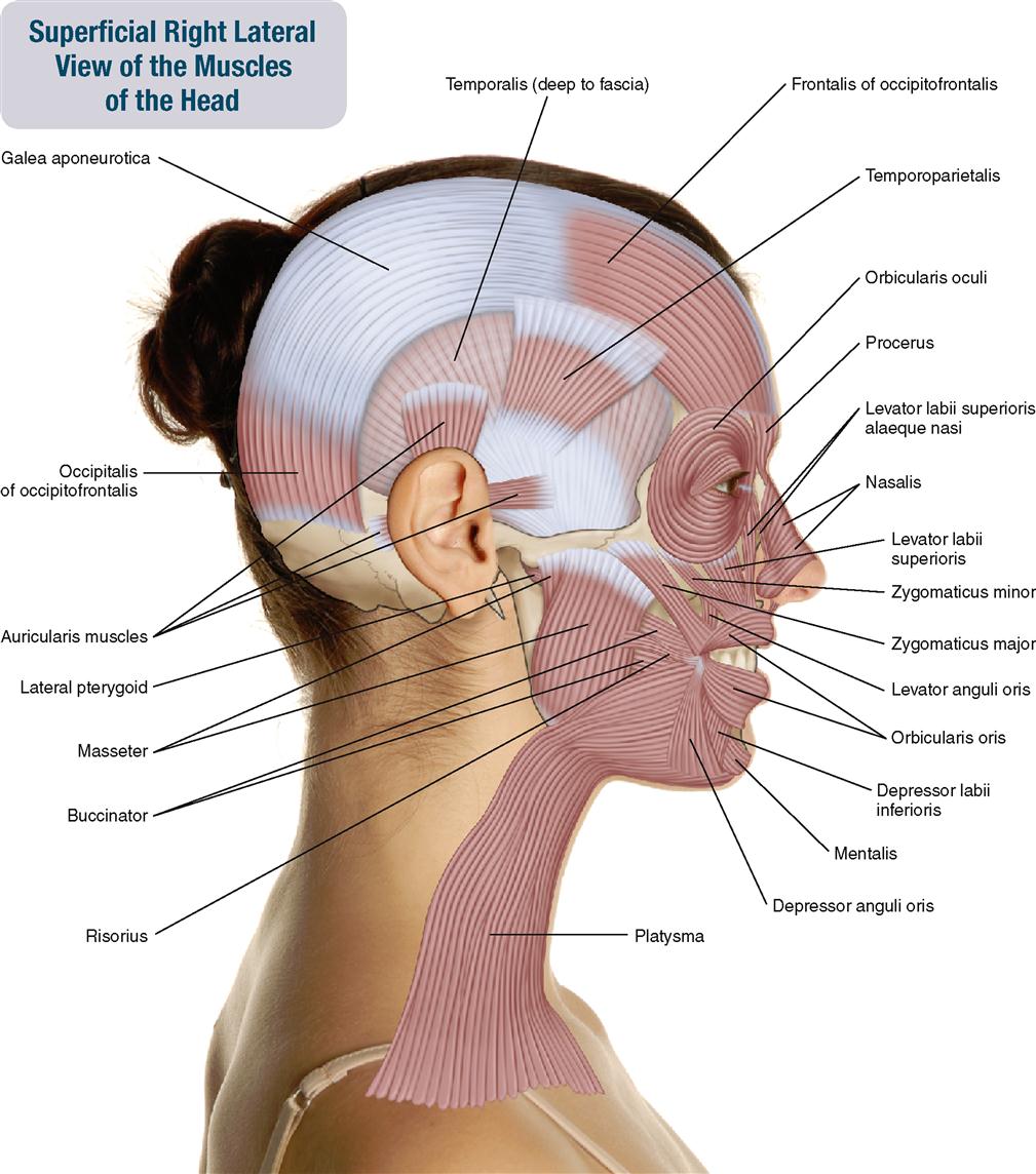
MUSCLES OF THE HEAD
Temporalis; Masseter
Pronunciation tem-po-RA-lis • MA-sa-ter
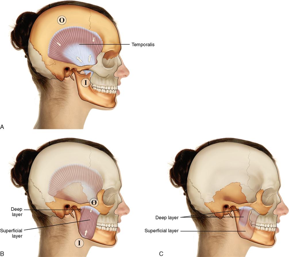
The temporalis and masseter are two of the four major muscles of mastication (chewing). The other two are the lateral and medial pterygoids discussed in the next layout. The temporalis and masseter are superficial muscles; the temporalis overlies the temporal bone and the masseter overlies the mandible (Figure 9-4). The masseter is usually divided into two layers: a superficial layer and a deep layer.
ATTACHMENTS
Temporalis
Origin (Superior Attachment)

Insertion (Inferior Attachment)

Masseter
Origin (Superior Attachment)

Insertion (Inferior Attachment)

ACTIONS
Both the temporalis and masseter move the mandible at the temporomandibular joints (TMJs).

STABILIZATION
Stabilize the mandible at the TMJs.
INNERVATION

PALPATION
Both the temporalis and masseter are palpated with the client supine.
Temporalis
1. Place your palpating finger pads over the temporal fossa on the head (superior to the ear).
2. Ask the client to alternately contract and relax the temporalis; clenching the teeth and then relaxing the jaw will accomplish this. Feel for the contraction of the temporalis as the client clenches the teeth (Figure 9-5).
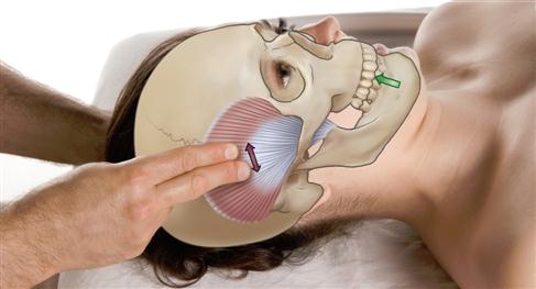
Masseter
1. Place your palpating finger pads between the zygomatic arch and the angle of the mandible.
2. Ask the client to alternately contract and relax the masseter; clenching the teeth and then relaxing the jaw will accomplish this. Feel for the contraction of the masseter as the client clenches the teeth (Figure 9-6).
TREATMENT CONSIDERATIONS


MUSCLES OF THE HEAD: Pterygoid Group
Lateral Pterygoid; Medial Pterygoid
Pronunciation LAT-er-al TER-i-goyd • MEE-dee-al TER-i-goyd
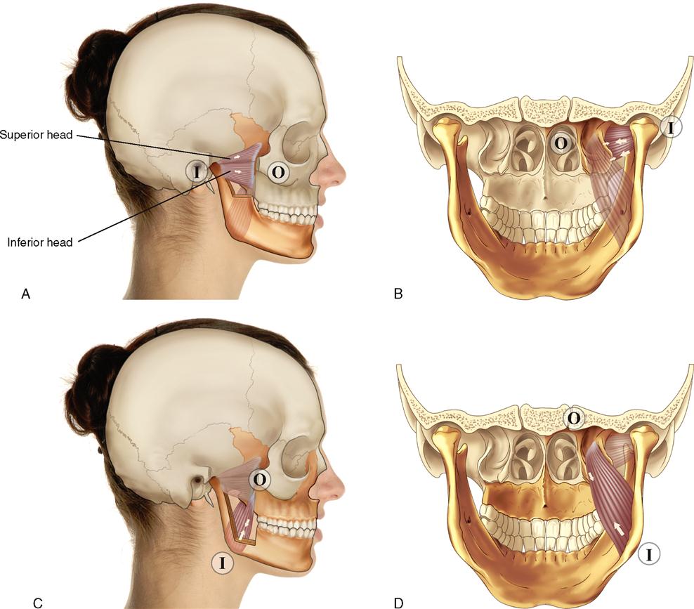
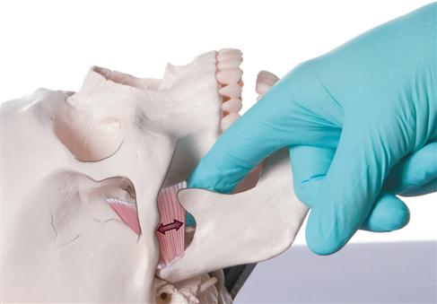
The pterygoid group is composed of the lateral pterygoid and medial pterygoid muscles (Figure 9-7). The pterygoids are two of the four major muscles of mastication (chewing). The temporalis and masseter are the other two muscles and are discussed in the previous layout. Both pterygoids are deep to the mandible and best accessed from inside the mouth. The lateral pterygoid has two heads: a superior head and an inferior head.
ATTACHMENTS
Lateral Pterygoid
Origin (Anterior/Medial Attachment)

Insertion (Posterior/Lateral Attachment)

Medial Pterygoid
Origin (Anterior/Medial/Superior Attachment)

Insertion (Posterior/Lateral/Inferior Attachment)

ACTIONS

Lateral Pterygoid


Medial Pterygoid


STABILIZATION
Stabilizes the mandible at the TMJs.
INNERVATION

PALPATION
Both pterygoids are palpated with the client supine.
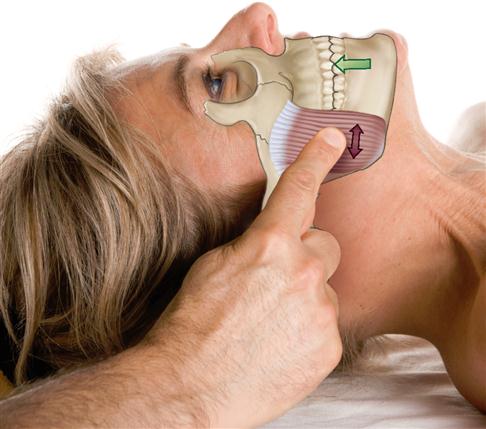
Lateral Pterygoid
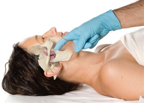
1. Wearing either a glove or a finger cot, place your palpating finger inside the vestibule of the client’s mouth (between the cheeks and the teeth) and run along the external surfaces of the upper teeth until you reach the back molars. Then press posteriorly and superiorly into a little pocket in the tissue between the gum above the upper teeth and the condyle of the mandible. You will be on the internal surface of the lateral pterygoid (Figure 9-8).
2. Now ask the client to gently either protract the mandible at the TMJs or deviate contralaterally the mandible slowly and carefully (deviate it to the opposite side of the body). Feel for the contraction of the lateral pterygoid (Figure 9-9).
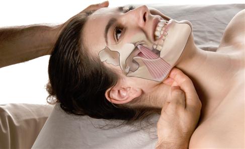
Medial Pterygoid
2. Now ask the client to protract the mandible. Feel for the contraction of the medial pterygoid.
3. Once felt, palpate as much of the medial pterygoid as possible.
5. Ask the client to elevate the mandible at the TMJs by clenching the teeth. Feel for the contraction of the medial pterygoid (Figure 9-10).
TREATMENT CONSIDERATIONS

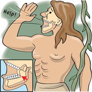
 A tight lateral or medial pterygoid may be involved with dysfunction of the TMJ (TMJ syndrome).
 The lateral pterygoid functions to protract the mandible and the articular disc of the TMJ. It is important that the mandible and disc protract together when the jaw is opened. Therefore if the contraction of the lateral pterygoid is not precisely coordinated with the other muscles that move the mandible, the articular disc may become jammed between the two bones of the joint and dysfunction of the TMJ (TMJ syndrome) may occur.
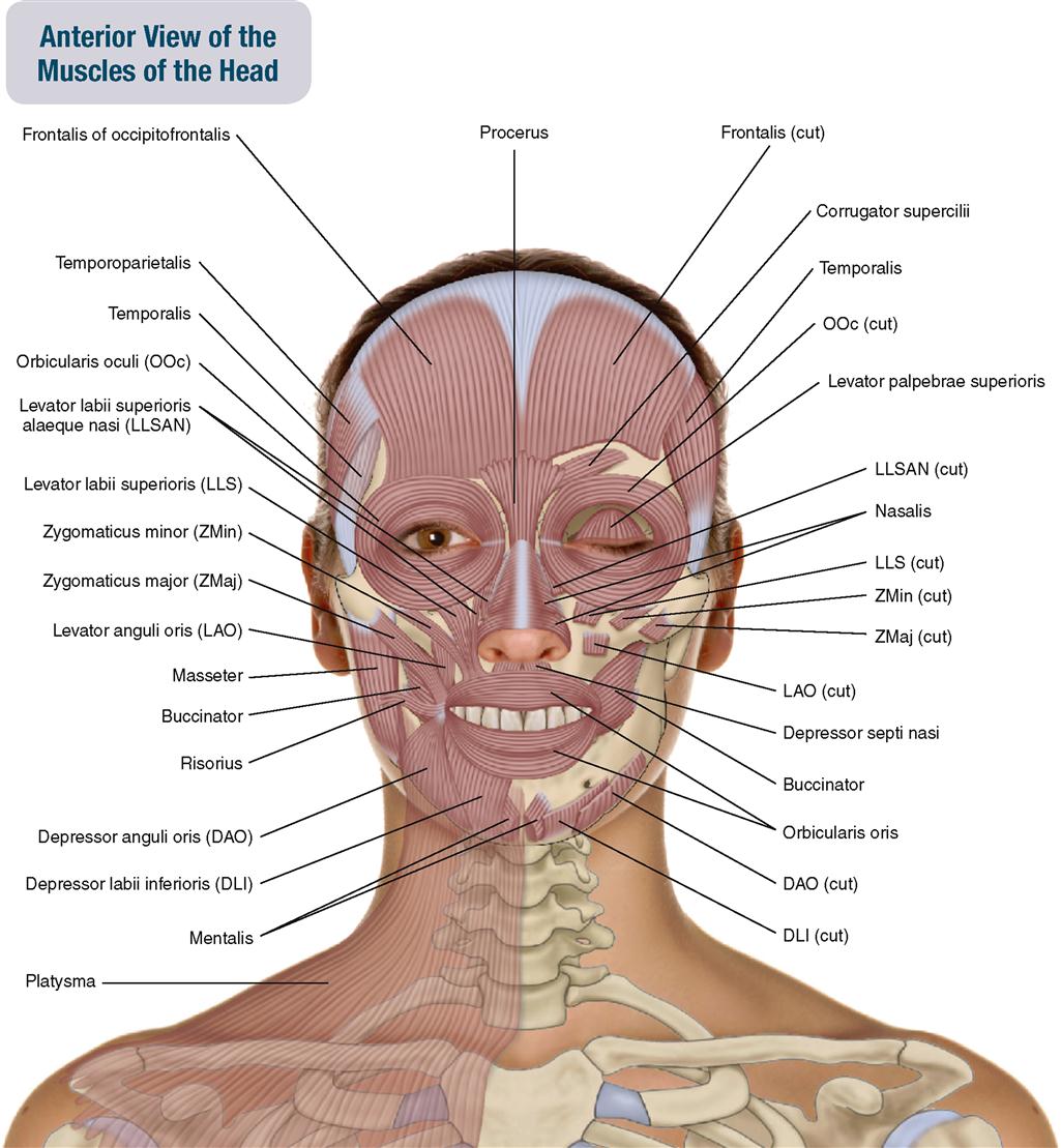
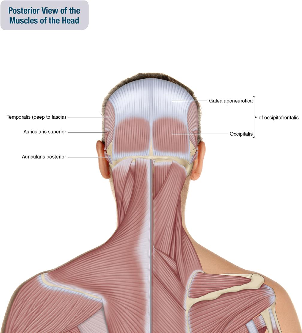
MUSCLES OF THE HEAD: Muscles of the Scalp
Occipitofrontalis; Temporoparietalis; Auricularis Anterior; Auricularis Posterior; Auricularis Superior
Pronunciation ok-SIP-i-to-fron-TA-lis • TEM-po-ro-pa-RI-i-TAL-is • aw-RIK-u-la-ris an-TEE-ri-or • aw-RIK-u-la-ris pos-TEE-ri-or • aw-RIK-u-la-ris sue-PEE-ri-or
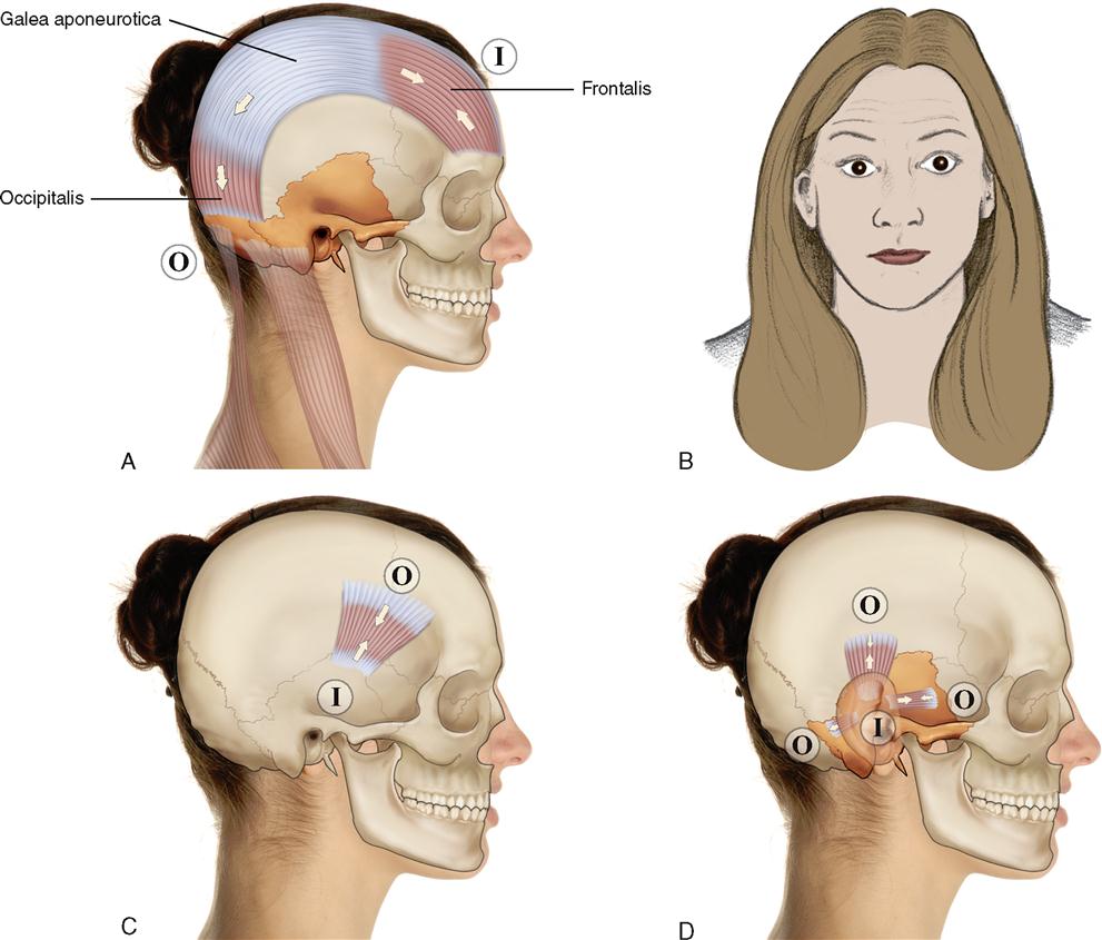
The muscles of the scalp are the occipitofrontalis (consisting of an occipitalis belly, a frontalis belly, and the intermediate galea aponeurotica), the temporoparietalis, and the three muscles of the auricularis group (auricularis anterior, auricularis posterior, and auricularis superior) (Figure 9-11). The occipitofrontalis and temporoparietalis are sometimes grouped together as the epicranius muscle. The muscles of the scalp are superficial fascial muscles.
ATTACHMENTS
Occipitofrontalis
Origin (Posterior Attachment)

Insertion (Anterior Attachment)

Temporoparietalis
Origin (Superior Attachment)

Insertion (Inferior Attachment)

Auricularis Group
Auricularis Anterior
Origin (Anterior Attachment)

Insertion (Posterior Attachment)

Auricularis Superior
Origin (Superior Attachment)

Insertion (Inferior Attachment)

Auricularis Posterior
Origin (Posterior Attachment)

Insertion (Anterior Attachment)

ACTIONS
Occipitofrontalis

Temporoparietalis

Auricularis Group


INNERVATION

PALPATION
All three muscles and muscle groups are palpated with the client supine.
Occipitofrontalis
1. Place your palpating finger pads on the forehead of the client.
2. Ask the client to elevate the eyebrows. Feel for the contraction of the frontalis belly (Figure 9-12, A); once felt, palpate the entire frontalis.
3. Now palpate over the client’s occipital bone, and ask the client to elevate the eyebrows. Feel for the contraction of the occipitalis belly (Figure 9-12, B); once felt, palpate the entire occipitalis.

Stay updated, free articles. Join our Telegram channel

Full access? Get Clinical Tree


