Muscles of the Forearm and Hand
The muscles of this chapter are involved with motions of the forearm (radius and ulna) at the radioulnar joints, the hand at the wrist (radiocarpal) joint, and the fingers at the metacarpophalangeal (MCP) and/or the proximal interphalangeal (PIP) and distal interphalangeal (DIP) joints; the thumb also moves at the first carpometacarpal (CMC) (saddle) joint.
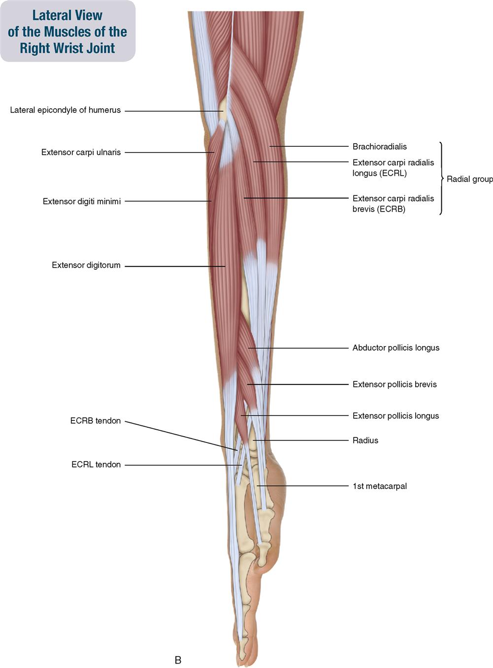
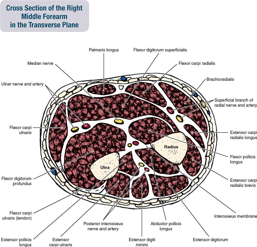
Forearm muscles are usually divided into an anterior flexor compartment and a posterior extensor compartment. The flexor compartment has three layers: superficial, intermediate, and deep. The extensor compartment has two layers: superficial and deep. A third group, called the radial group (also known as the wad of three), is sometimes designated. It consists of the brachioradialis of the anterior compartment and the extensors carpi radialis longus and brevis of the posterior compartment.
Two other structures of importance in the forearm are the common flexor tendon and the common extensor tendon. The common flexor tendon attaches to the medial epicondyle of the humerus. Five muscles attach into the common flexor tendon: (1) flexor carpi radialis, (2) palmaris longus, (3) flexor carpi ulnaris, (4) pronator teres, and (5) flexor digitorum superficialis. The common extensor tendon attaches to the lateral epicondyle of the humerus. Four muscles attach into the common extensor tendon: (1) extensor carpi radialis brevis, (2) extensor digitorum, (3) extensor digiti minimi, and (4) extensor carpi ulnaris.
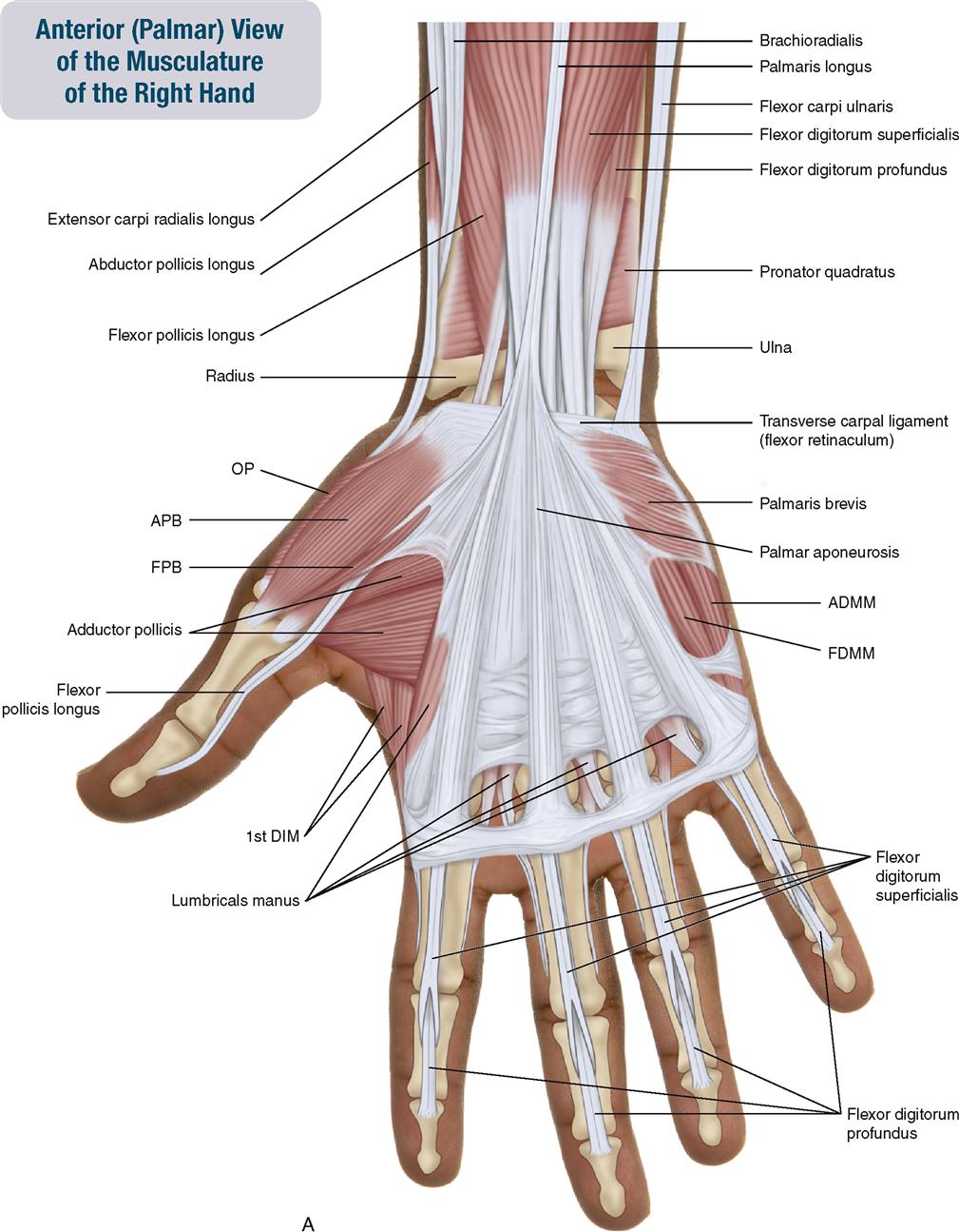
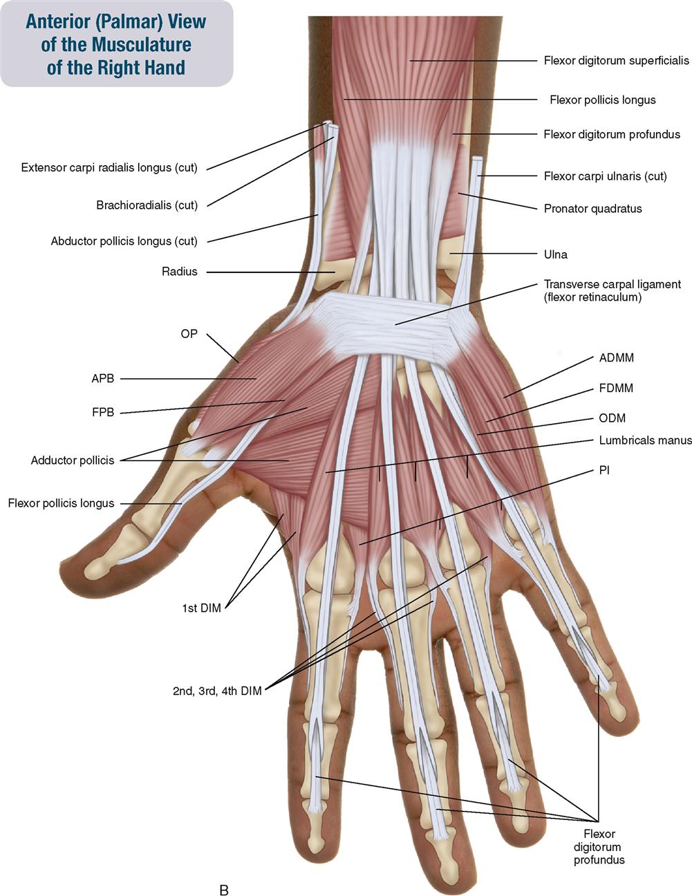
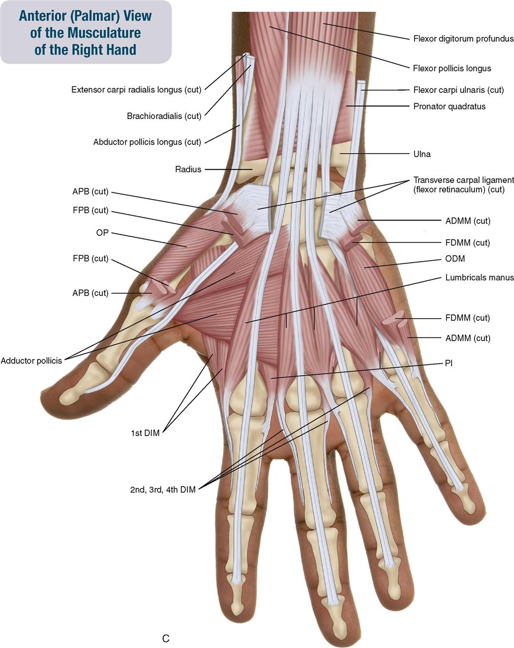
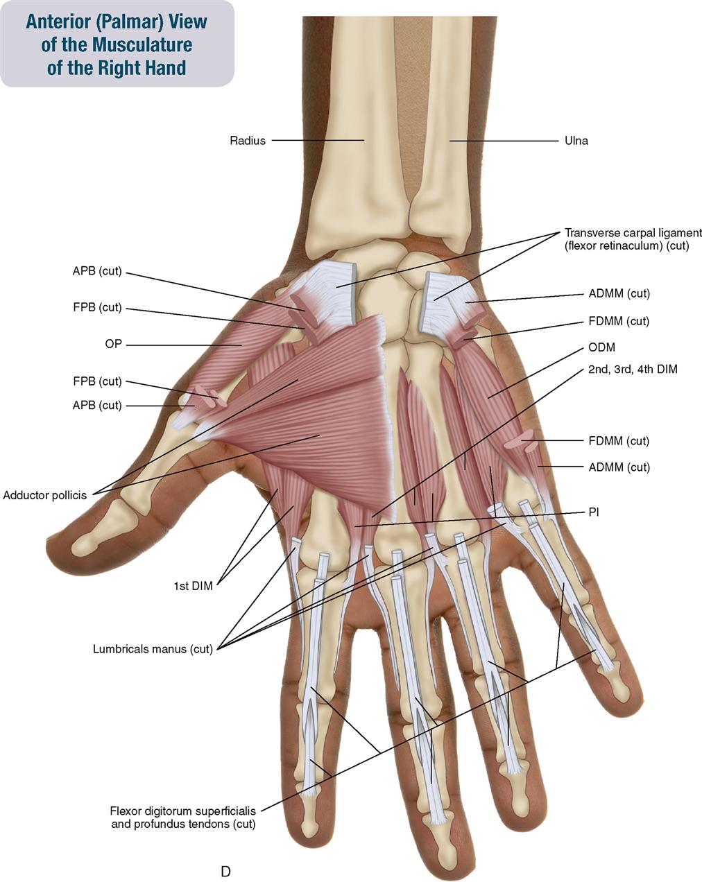
Muscles that move the fingers are often divided into extrinsic and intrinsic hand/finger muscles. Intrinsic hand muscles are wholly located within the hand; in other words, they originate and insert within the hand. Intrinsic muscles on the palmar side of the hand can be divided into three groups: (1) thenar eminence, (2) hypothenar eminence, and (3) central compartment.
Extrinsic finger muscles have their origin (proximal attachment) outside of the hand, in the forearm or arm. Because they also cross the wrist and/or elbow joints, they can also move those joints.
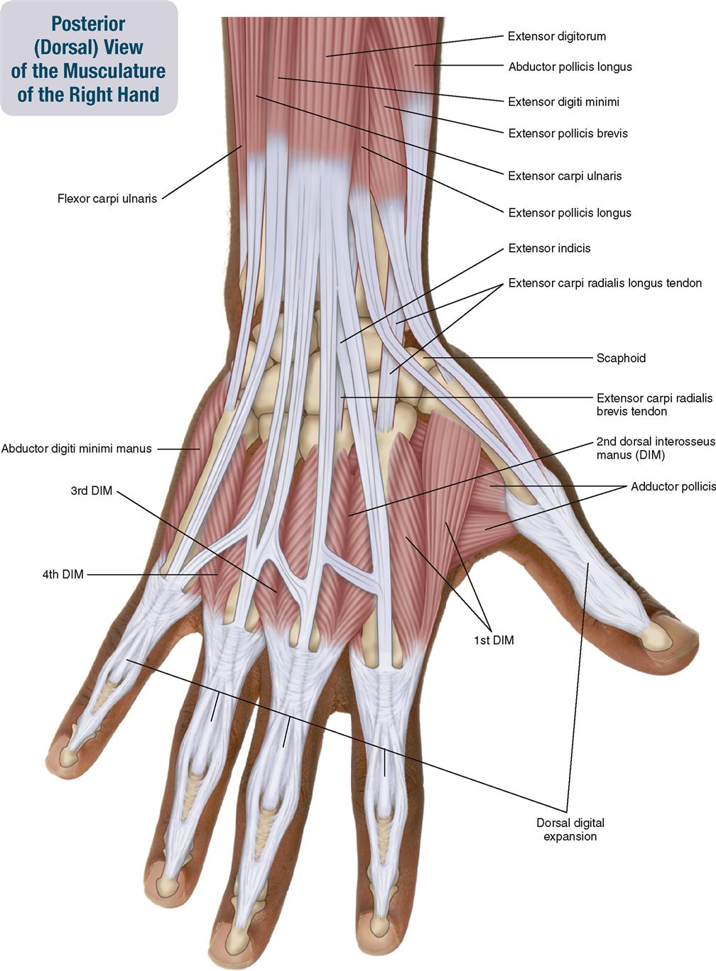
A structure of importance in the hand is the dorsal digital expansion. The dorsal digital expansion is a fibrous expansion of the extensor digitorum and extensor pollicis longus muscles’ distal tendons on the dorsal side of the fingers (digits).
As a general rule, muscles that move the elbow joint have their origin (proximal attachment) on the arm (humerus) and their insertion (distal attachment) on the forearm (radius or ulna) or hand. Muscles that pronate or supinate the forearm usually have their origin (proximal attachment) on the radius and their insertion (distal attachment) on the ulna. Muscles that move the wrist joint usually have their origin (proximal attachment) on the arm or forearm and their insertion (distal attachment) on the hand. Finger muscles may be extrinsic or intrinsic as previously discussed.
As a rule, flexor and pronator muscles attach to the medial epicondyle of the humerus via the common flexor tendon.
As a rule, extensor and supinator muscles attach to the lateral epicondyle of the humerus via the common extensor tendon.
The companion CD at the back of this book allows you to examine the muscles of this body region, layer by layer, and individual muscle palpation technique videos are available in the Chapter 7 folder on Evolve.
OVERVIEW OF FUNCTION: MUSCLES OF THE ELBOW AND RADIOULNAR JOINTS
The following general rules regarding actions can be stated for the functional groups of the muscles of the elbow and radioulnar joints.


 Reverse actions at the elbow joint involve moving the arm toward the forearm at the elbow joint. This movement usually occurs when the hand (and therefore the forearm) is fixed by holding onto an immovable object.*


 Reverse actions of these standard mover actions at the radioulnar joints involve moving the ulna toward the radius at the radioulnar joints. This movement usually occurs when the hand (and therefore the radius) is fixed by holding onto an immovable object.*
OVERVIEW OF FUNCTION: MUSCLES OF THE WRIST JOINT
The following general rules regarding actions can be stated for the functional groups of the muscles of the wrist joint:


OVERVIEW OF FUNCTION: MUSCLES OF THE FINGERS
The following general rules regarding actions can be stated for the functional groups of finger muscles:
 Fingers two through five can move at three joints: (1) MCP, (2) PIP, and (3) DIP joints. If a muscle crosses only the MCP joint, it can move the finger only at the MCP joint. If the muscle crosses the MCP and PIP joints, it can move the finger at both of these joints. If the muscle crosses the MCP, PIP, and DIP joints, it can move the finger at all three joints.


 Reverse actions involve the proximal attachment moving toward the distal attachment. This movement occurs when the fingers are holding onto a fixed, immovable object.*

Notes
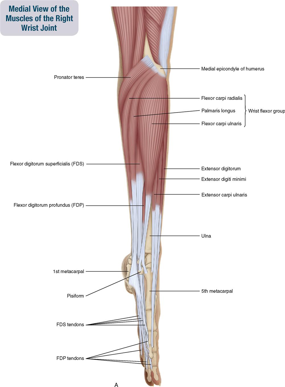
FOREARM AND HAND: Wrist Flexor Group
Flexor Carpi Radialis; Palmaris Longus; Flexor Carpi Ulnaris
Pronunciation FLEKS-or KAR-pie RAY-dee-A-lis • pall-MA-ris LONG-us • FLEKS-or KAR-pie ul-NA-ris
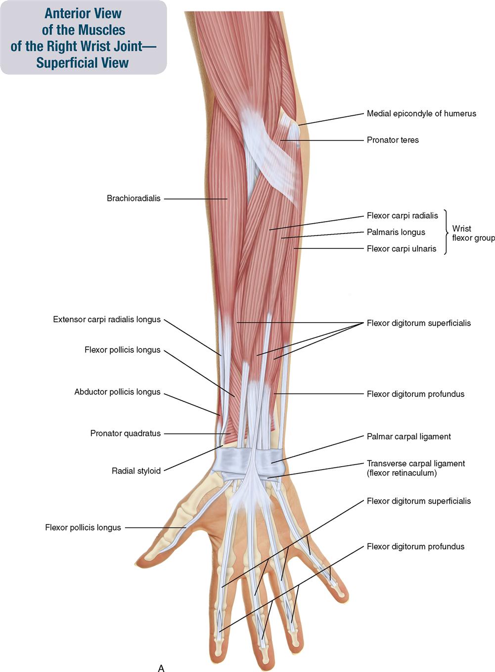
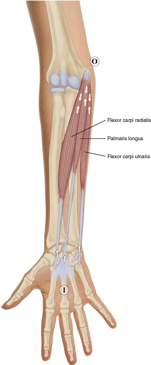
The wrist flexor group is composed of three muscles that all originate (have their proximal attachment) on the medial epicondyle of the humerus via the common flexor tendon. They all cross the wrist joint anteriorly; therefore they can all flex the hand at the wrist joint, hence the name of the group. These muscles are the flexor carpi radialis, palmaris longus, and flexor carpi ulnaris. All three muscles of the wrist flexor group are superficial in the anterior forearm. At the wrist joint, the palmaris longus crosses dead center; the flexor carpi radialis crosses slightly to the radial (lateral) side; and the flexor carpi ulnaris crosses far to the ulnar (medial) side (Figure 7-7). In addition to the humeral attachment, the flexor carpi ulnaris also has an ulnar attachment. The humeral head is much thicker; the ulnar head is extremely thin.
ATTACHMENTS
Flexor Carpi Radialis
The flexor carpi radialis is superficial in the anterior forearm and located between the pronator teres and the palmaris longus.
Origin (Proximal Attachment)

Insertion (Distal Attachment)

Palmaris Longus
The palmaris longus is superficial in the anterior forearm and located between the flexor carpi radialis and the flexor carpi ulnaris.
Origin (Proximal Attachment)

Insertion (Distal Attachment)

Flexor Carpi Ulnaris
The flexor carpi ulnaris is superficial in the anterior forearm and located medial to the palmaris longus.
Origin (Proximal Attachment)

Insertion (Distal Attachment)

ACTIONS
 All three muscles of the wrist flexor group flex the hand at the wrist joint.
 The flexor carpi radialis also radially deviates the hand at the wrist joint.
 The flexor carpi ulnaris also ulnar deviates the hand at the wrist joint.
STABILIZATION
As a group, the wrist flexor muscles stabilize the wrist, elbow, and radioulnar joints.
INNERVATION

PALPATION
3. Begin by palpating the flexor carpi radialis by strumming horizontally across it (Figure 7-8). Then palpate the palmaris longus and flexor carpi ulnaris in a similar manner.
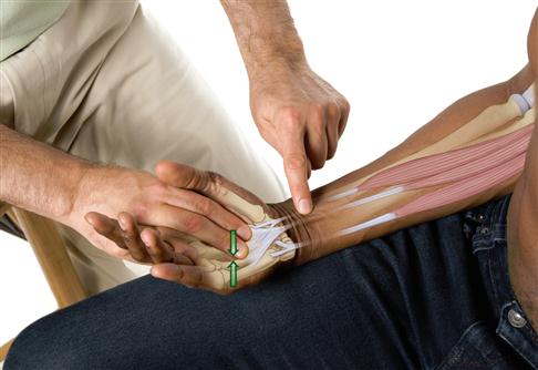
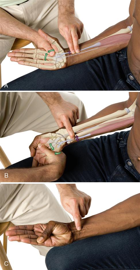
Note: The flexor carpi radialis can be palpated with resisted radial deviation of the hand at the wrist joint; similarly, the flexor carpi ulnaris can be palpated with ulnar deviation. The palmaris longus can be palpated by asking the client to “cup the hand” (Figure 7-9).
TREATMENT CONSIDERATIONS
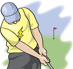
 Overuse of the wrist flexor group musculature can cause irritation and/or inflammation of the medial epicondyle and/or the common flexor tendon. This condition is known as medial epicondylitis, medial epicondylosis, or golfer’s elbow
 In many individuals, the palmaris longus is bilaterally or unilaterally absent.
 The ulnar nerve passes between the two heads of the flexor carpi ulnaris. Compression of the ulnar nerve between the two heads of the flexor carpi ulnaris is called cubital tunnel syndrome.
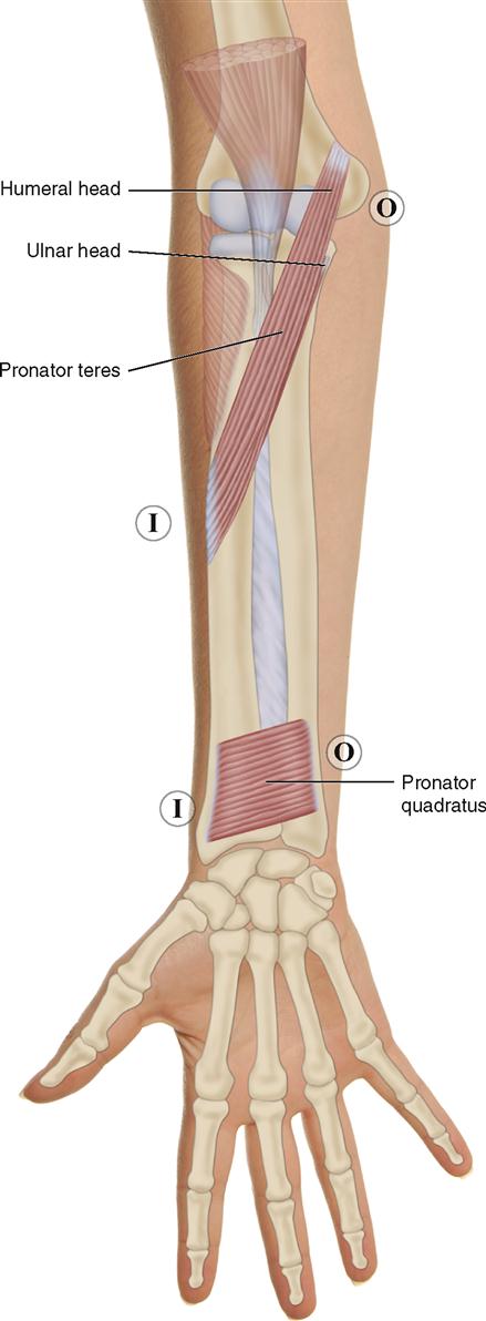
FOREARM AND HAND: PRONATOR GROUP
Pronator Teres; Pronator Quadratus
Pronunciation pro-NAY-tor TE-reez • pro-NAY-tor kwod-RAY-tus
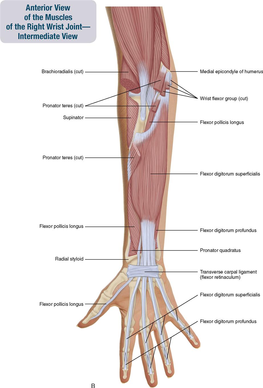
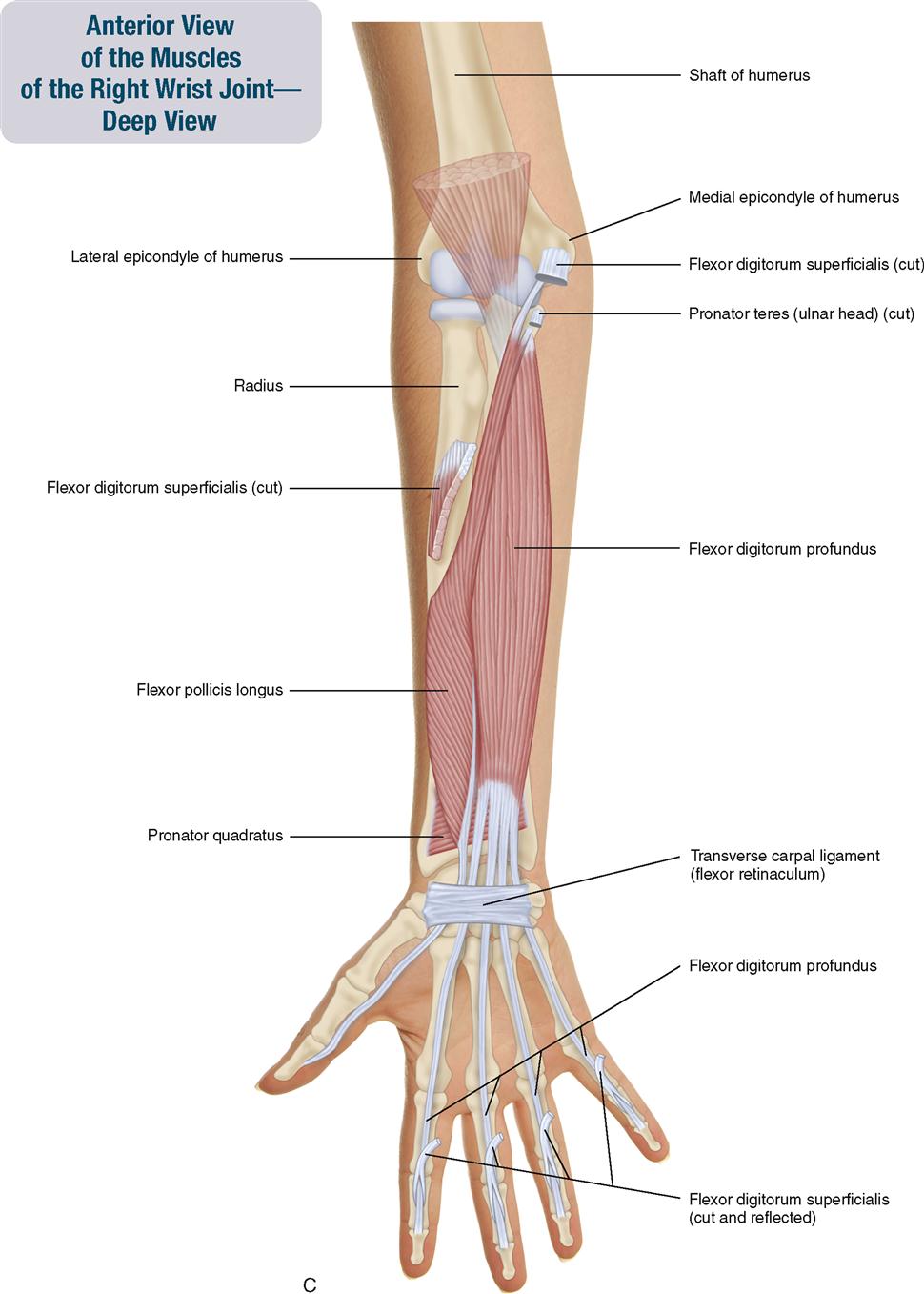
The pronator teres and pronator quadratus muscles are located in the anterior forearm. The pronator teres is superficial in the proximal forearm; the pronator quadratus is very deep in the distal forearm. The pronator teres has two heads—a large, superficial humeral head and a small, deep ulnar head (Figure 7-10).
ATTACHMENTS
Pronator Teres
Origin (Proximal Attachment)


Insertion (Distal Attachment)

Pronator Quadratus
Origin (Proximal Attachment)

Insertion (Distal Attachment)

ACTIONS
Pronator Teres


Pronator Quadratus

STABILIZATION
INNERVATION

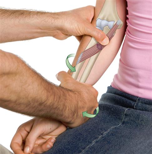
PALPATION
Pronator Teres
2. With moderate force, resist the client from pronating the forearm at the radioulnar joints and feel for the contraction of the pronator teres (Figure 7-11).
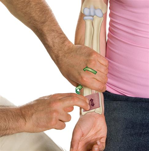
Pronator Quadratus
2. Ask the client to pronate the forearm actively at the radioulnar joints, and feel for the contraction of the pronator quadratus. Resistance can be added if necessary (Figure 7-12).
TREATMENT CONSIDERATIONS


FOREARM AND HAND
Brachioradialis
Pronunciation BRAY-key-o-RAY-dee-AL-is
The brachioradialis is superficial for its entire course (except for a small part of its distal tendon that is deep to two small muscles of the thumb whose bellies are located deep in the posterior forearm). The brachioradialis is located in the anterior forearm on the radial (lateral) side. Because it is on the radial side, it is considered to be part of the “radial group,” along with the extensor carpi radialis longus and extensor carpi radialis brevis (Figure 7-13).
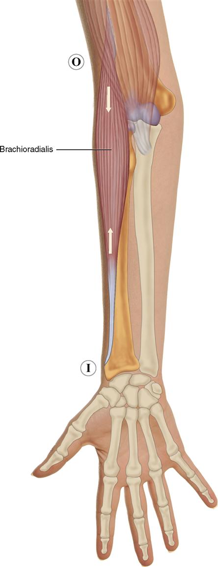
ATTACHMENTS
Origin (Proximal Attachment)

Insertion (Distal Attachment)

ACTIONS
The brachioradialis moves the forearm at the elbow and radioulnar joints.


STABILIZATION
Stabilizes the elbow and radioulnar joints.
INNERVATION

PALPATION
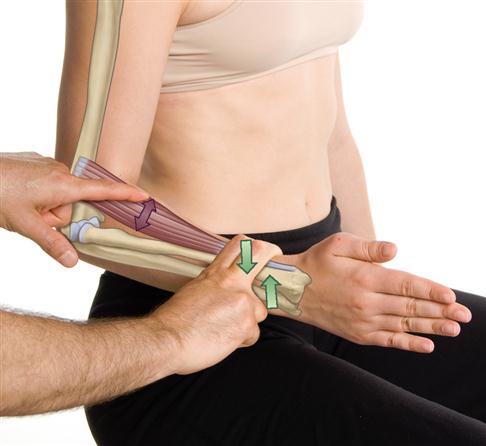
2. Ask the client to try to flex the forearm with moderate force against your resistance. First look for the contraction of the brachioradialis and then feel for its contraction at the proximal anterolateral forearm (Figure 7-14).
TREATMENT CONSIDERATIONS


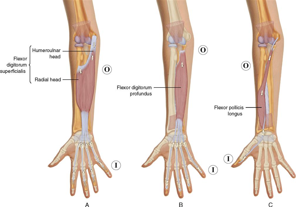
FOREARM AND HAND: Flexors Digitorum and Pollicis Group
Flexor Digitorum Superficialis: Flexor Digitorum Profundus; Flexor Pollicis Longus
Pronunciation FLEKS-or dij-i-TOE-rum SOO-per-fish-ee-A-lis •
FLEKS-or dij-i-TOE-rum pro-FUN-dus • FLEKS-or POL-i-sis LONG-us
The flexor digitorum superficialis, flexor digitorum profundus, and flexor pollicis longus are all long, extrinsic flexors of the fingers. Both flexor digitorum muscles flex fingers 2 through 5; and the flexor pollicis muscle flexes the thumb (finger 1). These muscles are considered to be long extrinsic finger muscles because they originate (have their proximal attachment) outside of the hand. The flexor digitorum superficialis is in the intermediate layer of anterior forearm muscles, directly deep to the muscles of the wrist flexor group. The flexor digitorum profundus and flexor pollicis longus are in the deep layer of the anterior forearm, deep to the flexor digitorum superficialis (Figure 7-15).
ATTACHMENTS
Flexor Digitorum Superficialis
Origin (Proximal Attachment)

Insertion (Distal Attachment)

Flexor Digitorum Profundus
Origin (Proximal Attachment)

Insertion (Distal Attachment)

Flexor Pollicis Longus
Origin (Proximal Attachment)


Stay updated, free articles. Join our Telegram channel

Full access? Get Clinical Tree


