• The abdomen must be kept free, and bolsters are placed at the chest and iliac crest regions. • The arms are flexed at the elbows and placed forward with the axilla remaining free of any compression. • The head needs to be placed in a standard headrest with access at the cervicothoracic junction.
Posterior Surgical Treatment for Scheuermann’s Kyphosis
Examination/Imaging
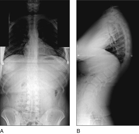
 Standing posteroanterior (PA) and lateral 36-inch radiographs are obtained. Figure 1 shows standing radiographs of a patient with kyphosis in PA (Fig. 1A) and lateral (Fig. 1B) views.

 A lateral radiograph in hyperextesion with a bolster under the kyphotic apex is also obtained.
 Magnetic resonance imaging is done to assess for any possible thoracic disk herniations.
Surgical Anatomy
 Posterior-only treatment for Scheuermann’s kyphosis requires an understanding of the posterior anatomic structures of the spine as well as the variations in pedicle anatomy.
 Intimate knowledge of the posterior structures allows for the surgeon to easily perform Ponte osteotomies (foramina to foramina) over the apex of the curve.
Positioning
 The patient may be placed prone on a standard radiolucent spinal operating frame.
 The patient must have appropriate somatosensory and motor evoked potential monitoring in place.
Portals/Exposures
 The exposure of the thoracic and lumbar spine entails standard techniques.
 Hypotensive anesthesia is useful, taking care to keep the mean arterial pressures in the 65- to 70-mm Hg range.
 Subperiosteal dissection is necessary in order to obtain adequate bone for fusion as well as clear anatomic landmarks for pedicle screw placement.
 Exposure is done with Cobb elevators and electrocautery, taking care to expose out to the tips of the spinous processes.
Procedure
Step 1
 Pedicle screw placement begins distally in the lumbar spine, taking care to include the lumbar vertebra that is centered over the sacral spine in the lateral view.
 The use of fixed or variable screws will lead to reliable results in correcting kyphosis. The use of reduction screws at the distal levels may help capture the rods more easily, and may aid in rod capture proximally as well.
 Standard lumbar screw placement is performed, taking care to place as large a diameter of screw as possible, with the length of screws typically ranging from 40 to 50 mm.
 Thoracic screw placement is done after facetectomies at each corresponding level.
![]()
Stay updated, free articles. Join our Telegram channel

Full access? Get Clinical Tree


56: Posterior Surgical Treatment for Scheuermann’s Kyphosis


