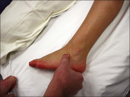
• The patient is observed for an antalgic gait, characterized by a decreased stance phase on the affected extremity, and the position of the foot during stance phase is evaluated. • Hindfoot and midfoot alignment are examined. Patients with a calcaneonavicular coalition will typically present with hindfoot valgus and abduction across the midfoot. • The patient is evaluated for flattening of the medial longitudinal arch. • A single-leg heel rise will assess the flexibility of the flatfoot deformity. Failure to reconstitute the arch of the foot is indicative of a rigid hindfoot and suggests the presence of some abnormality within the subtalar or transverse tarsal joints, such as a tarsal coalition. • In a calcaneonavicular coalition, point tenderness may be elicited directly over and distal to the anterior process of the calcaneus (Fig. 1). • The hindfoot is inverted and everted while stabilizing the ankle joint, and compared to the contralateral side. Tarsal coalitions are associated with pain and limitation of subtalar motion. • Anteroposterior (AP), lateral, and oblique views of the foot are obtained. Figure 2A shows an AP view of child with a calcaneonavicular coalition. • The oblique and lateral views of the foot are evaluated. • Standing AP, lateral, and Saltzman hindfoot alignment views may be used to assess alignment.
Resection of Calcaneonavicular Coalition and Fat Autograft Interposition
Examination/Imaging
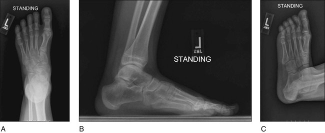
 The “anteater nose” sign is a prominent anterior process of the calcaneus seen on the lateral view (Fig. 2B).
 The oblique view is the best for visualization of a calcaneonavicular coalition. In Figure 2C, note the connection between the anterior process of the calcaneus and the lateral aspect of the navicular
 CT scan is necessary evaluate for concomitant talocalcaneal coalitions.
Positioning
 The patient is placed supine on a radiolucent operating room table (Fig. 3).
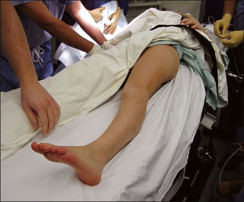
 A bump is placed under the hip of the operative leg to internally rotate the extremity.
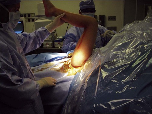
 The extremity is prepped up to the buttocks to allow for harvesting of the fat autograft. The drapes are placed as proximal as possible to ensure that there is adequate visualization of the gluteal crease (Fig. 4).
Portals/Exposures
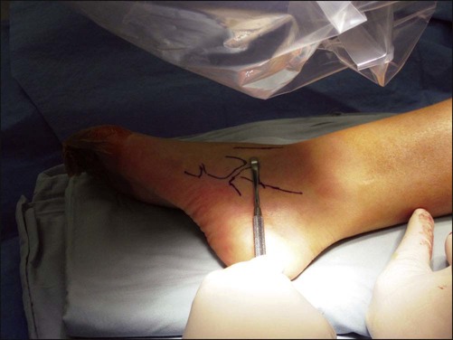
 Landmarks are drawn on the skin (Fig. 5). The anterior process of the calcaneus, the cuboid, the fifth metatarsal, and the peroneal and extensor tendons, are marked. An elevator is used with fluoroscopy to find the correct level directly above the coalition.
 The incision is marked on the skin. It lies between the extensor and peroneal tendons (Fig. 6).
Related posts:
 4: Open Reduction and Internal Fixation of Displaced Medial Epicondyle Fracture Using a Screw and Washer
29: Epiphysiodesis of the Distal Femur/Proximal Tibia-Fibula
32: Patellar Instability: Lateral Release and Medial Plication
31: Discoid Lateral Meniscus
39: Open Reduction and Internal Fixation of Tibial Tubercle Fractures
10: Digital Syndactyly Release
4: Open Reduction and Internal Fixation of Displaced Medial Epicondyle Fracture Using a Screw and Washer
29: Epiphysiodesis of the Distal Femur/Proximal Tibia-Fibula
32: Patellar Instability: Lateral Release and Medial Plication
31: Discoid Lateral Meniscus
39: Open Reduction and Internal Fixation of Tibial Tubercle Fractures
10: Digital Syndactyly Release
![]()
Stay updated, free articles. Join our Telegram channel

Full access? Get Clinical Tree


47: Resection of Calcaneonavicular Coalition and Fat Autograft Interposition


