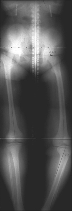
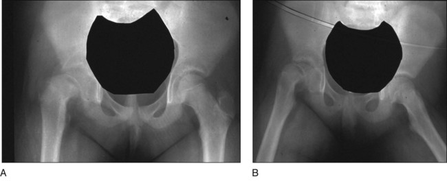
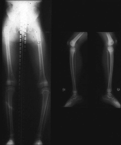
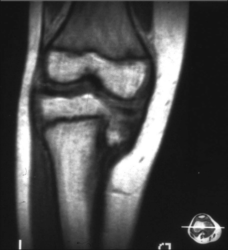
• Presence/absence of rotational abnormality • Hip examination for possible slipped capital femoral epiphysis (SCFE) • An orthoroentgenogram is obtained to assess alignment, ± femoral deformity, and limb length discrepancy. Figure 1 shows a typical preoperative orthoroentgenogram, obtained with the patient facing anterior, delineating coronal tibial and/or femoral deformity and limb length discrepancy. • Anteroposterior (AP) and frog-leg lateral radiographs are obtained to assess the hips. In Figure 2, the AP (Fig. 2A) and frog-leg lateral (Fig. 2B) radiographs demonstrate a right SCFE. • An AP/lateral radiograph is obtained of the entire tibia, including the ankle joint. Figure 3 shows an orthoroentgenogram and lateral radiograph of the tibia in a 6-year-old. • Computed tomography (CT) or magnetic resonance imaging (MRI) can be done if the patient is young or in a patient with long-standing problems to assess the joint surface. Figure 4 shows an MRI of the right proximal tibia delineating both the joint contour and the proximal physis. • CT or MRI is also used for possible growth plate assessment.
Proximal Tibial Osteotomy for Blount’s Disease
Examination/Imaging
 Complete physical examination with emphasis on:
Positioning
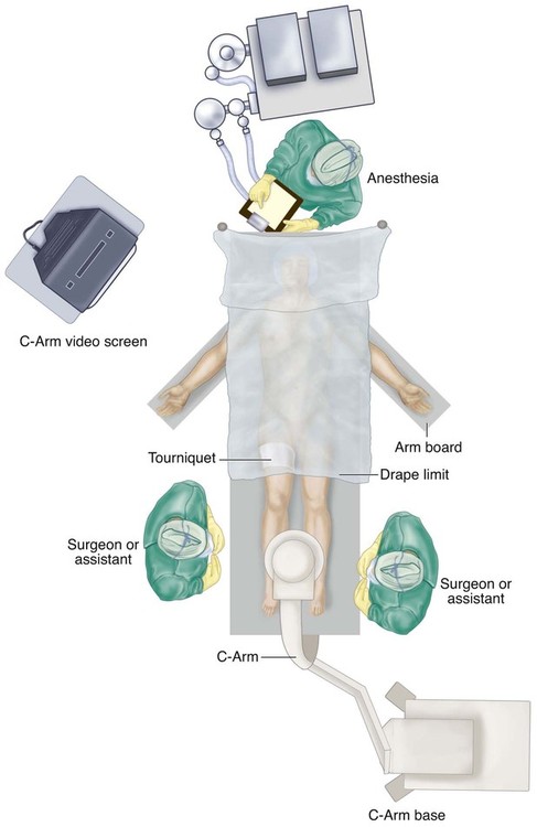
 The patient is placed supine with the patellae anterior and a tourniquet on the thigh. Draping is just distal to the tourniquet, leaving both legs visible for comparison, especially in acute correction (Fig. 5).
 The involved leg is prepped distal to the tourniquet.
 The C-arm is placed perpendicular to the end of the bed, allowing both the surgeon and the assistant access from either side, as shown in Figure 5.
Procedure: Small Wire Circular Fixators
Step 1
 Preoperative construction is based on patient size (ring size choice) and radiographs.
 The initial wire (when using wire fixators) should be smooth (Fig. 6
Related posts:
 4: Open Reduction and Internal Fixation of Displaced Medial Epicondyle Fracture Using a Screw and Washer
29: Epiphysiodesis of the Distal Femur/Proximal Tibia-Fibula
32: Patellar Instability: Lateral Release and Medial Plication
31: Discoid Lateral Meniscus
39: Open Reduction and Internal Fixation of Tibial Tubercle Fractures
10: Digital Syndactyly Release
4: Open Reduction and Internal Fixation of Displaced Medial Epicondyle Fracture Using a Screw and Washer
29: Epiphysiodesis of the Distal Femur/Proximal Tibia-Fibula
32: Patellar Instability: Lateral Release and Medial Plication
31: Discoid Lateral Meniscus
39: Open Reduction and Internal Fixation of Tibial Tubercle Fractures
10: Digital Syndactyly Release
![]()
Stay updated, free articles. Join our Telegram channel

Full access? Get Clinical Tree


40: Proximal Tibial Osteotomy for Blount’s Disease


