• The patella should glide between one and two quadrants medially and laterally. • If the patella glides one quadrant or less medially, the lateral retinaculum is tight. If it glides more than two quadrants medially, the lateral retinaculum is loose.
Patellar Instability
Lateral Release and Medial Plication
Examination/Imaging
 A quadrant glide test with the knee in 30° of flexion should be performed to check for lateral tightness.
 Standard views as well as sunrise views of both knees should be obtained prior to operating. The sunrise radiograph in Figure 1A reveals lateral tilt of the patella.
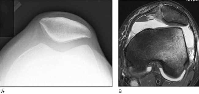
 Magnetic resonance imaging (MRI) can be helpful in determining whether chondral injury is present in the patellofemoral joint. An axial MRI after patellar dislocation (Fig. 1B) shows hemarthrosis, lateral subluxation of the patella, medial retinacular tear, lateral femoral condyle bone bruise, and patellar chondromalacia.
Surgical Anatomy
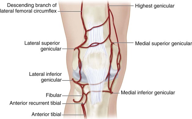
 The superior genicular vessels are near the vastus lateralis musculotendinous junction and must be coagulated thoroughly after lateral release to prevent hemarthrosis.
 Figure 2 shows the arterial supply to the knee. Note where the superolateral genicular artery nears the vastus lateralis.
Portals/Exposures
 A standard anterolateral portal should be made next to the patellar tendon and at the level of the inferior pole of the patella.
 An anteromedial portal is made in the standard position.
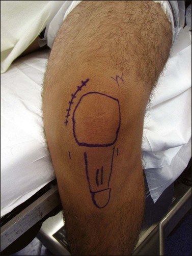
 Medial plication requires a 4-cm incision placed 1 cm medial to the medial border of the patella.
 Figure 3 shows the standard positions for arthroscopic portals, and the potential incision for a medial plication is marked.
Procedure: Arthroscopic Lateral Release
Step 1
 The anterolateral portal is made first, and a standard diagnostic arthroscopy proceeds. Patellar tilt and tracking should be observed in relation to the trochlear groove.
 The lateral release can be performed with either electrocautery or Metzenbaum scissors.
Related posts:
 4: Open Reduction and Internal Fixation of Displaced Medial Epicondyle Fracture Using a Screw and Washer
29: Epiphysiodesis of the Distal Femur/Proximal Tibia-Fibula
28: Femur Fracture: Closed Reduction and Spica Cast
46: Resection of Talocalcaneal Tarsal Coalition and Fat Autograft Interposition
57: Scoliosis Correction
10: Digital Syndactyly Release
4: Open Reduction and Internal Fixation of Displaced Medial Epicondyle Fracture Using a Screw and Washer
29: Epiphysiodesis of the Distal Femur/Proximal Tibia-Fibula
28: Femur Fracture: Closed Reduction and Spica Cast
46: Resection of Talocalcaneal Tarsal Coalition and Fat Autograft Interposition
57: Scoliosis Correction
10: Digital Syndactyly Release
![]()
Stay updated, free articles. Join our Telegram channel

Full access? Get Clinical Tree


Musculoskeletal Key
Fastest Musculoskeletal Insight Engine


