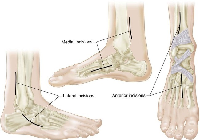
Tibialis Posterior Tendon Transfer
Indications
 Spastic, flexible equinovarus-cavovarus foot deformity in the ambulatory child or adult with cerebral palsy or brain injury with “over-pull” of the tibialis posterior.
 Recurrence of clubfoot deformity.
 Patients with weak dorsiflexion secondary to neuromuscular disease.
 Lengthening and transfer of the tendon have both been described.
 Additionally, one may transfer all or half of the tendon.
 Consideration should be given to whether the foot and ankle have adequate motor units to power dorsiflexion and are struggling with the varus over-pull of the tibialis posterior, in which case a split transfer might be advised; or the patient would benefit more from assistance with dorsiflexion and transfer of the entire tendon due to a general weakness of dorsiflexion.
Examination/Imaging
 The peroneus brevis may be relatively weaker, allowing the tibialis posterior to “over-pull” the midfoot into supination and the hindfoot into varus, especially during the swing phase of gait.
 The gastrocnemius-soleus must also be assessed for contracture.
 The goal is a balanced foot in both stance and gait.
 Hindfoot varus flexibility may be tested with the Coleman block test (Coleman and Chestnut, 1977).
 Preoperative plain films, including standing anteroposterior, lateral, and oblique views of both feet, should be obtained to confirm no fixed bony deformity.
Surgical Anatomy
 The tibialis posterior has a broad insertion and is best found proximal to the first ray through the medial incision.
 When transferring to the peroneus brevis, care should be taken not to completely disrupt the lateral fascia protecting the peroneal tendons from subluxation over the lateral malleolus.
Procedure
Step 1
 The tourniquet may be used at the surgeon’s discretion. The procedure does not typically result in a particularly bloody field.
 If a gastrocnemius-soleus lengthening is required, this should be performed first and the wound closed before proceeding with the tendon transfer portion of the surgery.
51: Tibialis Posterior Tendon Transfer


