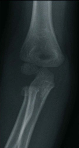
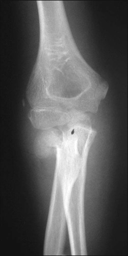
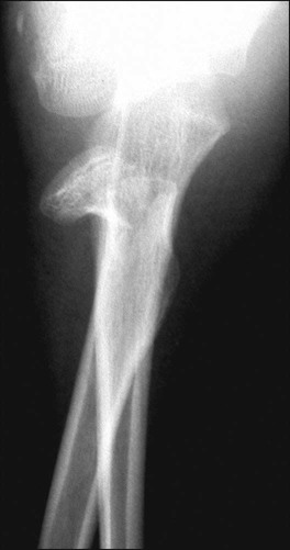
Sameer Badarudeen, Robert M. Bernstein and Saul M. Bernstein • Figure 1 is a radiograph showing fracture of the neck of the radius that could be treated with closed reduction or percutaneous reduction. • Figures 2 and 3 are radiographs of a fracture of the neck of the radius with complete dislocation that may be difficult to reduce closed. Equipment
Radial Head/Neck Fracture
Closed Reduction, Percutaneous Reduction, and Open Reduction
Examination/Imaging
 Neurovascular examination (posterior interosseous nerve)
 Orthogonal radiographs of the radial head and elbow joint
 Arthrogram in children in whom the radial head secondary center has not yet ossified
Surgical Anatomy
 The radial head and neck are subcutaneous on the lateral side of the elbow, just distal to the lateral epicondyle.
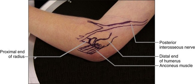
 The posterior interosseous nerve penetrates the supinator muscle just distal to the radial neck and may lie directly upon the bone (Fig. 4).
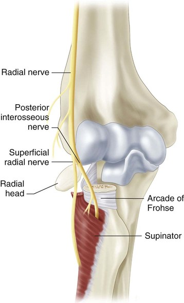
 Figure 5 depicts the surface anatomy of the posterior interosseous nerve in relation to the radial neck.
Closed Reduction
Positioning
 The patient is placed in the supine position on the table with the armboard at the ipsilateral head, folded in, to act as a headboard extension.
 The elbow is positioned over the fluoroscopic receiver (C-arm) (Fig. 6).
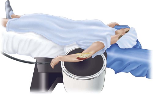
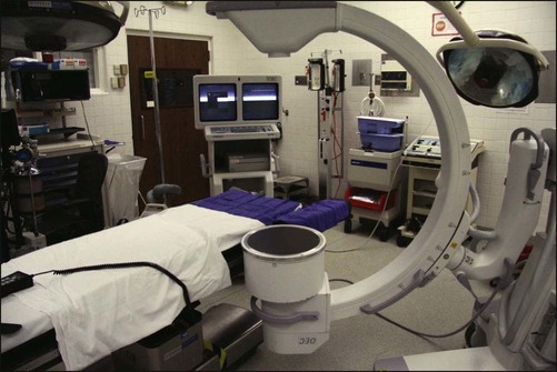
 Figure 7 depicts the authors’ preferred set up of the operating room for a “left” radial neck fracture, with the armboard and C-arm on the same side and the monitor on the opposite side of the table.
Related posts:
![]() 41: Operative Treatment of Tillaux Fractures of the Ankle
41: Operative Treatment of Tillaux Fractures of the Ankle
 7: Forearm Fractures: Closed Treatment
![]() 32: Patellar Instability: Lateral Release and Medial Plication
32: Patellar Instability: Lateral Release and Medial Plication
![]() 1: Modified Woodward Procedure for Sprengel’s Deformity
1: Modified Woodward Procedure for Sprengel’s Deformity
 40: Proximal Tibial Osteotomy for Blount’s Disease
 60: Thoracoscopic Release and Instrumentation for Scoliosis
![]()
Stay updated, free articles. Join our Telegram channel

Full access? Get Clinical Tree


5: Radial Head/Neck Fracture: Closed Reduction, Percutaneous Reduction, and Open Reduction


