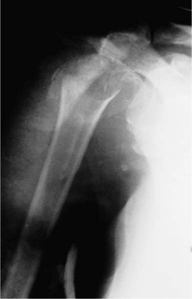
Case 5 A 73-year-old woman presents several hours after an injury sustained during a fall on her outstretched arm. She reports poor function and moderate to severe pain since the injury. She denies any numbness or weakness in the upper extremity but has limited motion. Range of motion is limited to 10 degrees of forward flexion and no external rotation. She is neurovascularly intact, with a strong 2+ radial and ulnar pulse. Her axillary nerve sensation is intact as well. She has moderate swelling about the shoulder and painful crepitus to any range of motion. Figure 5–1. An anteroposterior (AP) radiograph of the affected shoulder. 1. Proximal humerus fracture 2. Fracture-dislocation of the proximal humerus 3. Shoulder dislocation 4. Rotator cuff tear An anteroposterior (AP) radiograph of the shoulder was obtained (Fig. 5–1). PITFALLS • Failure to obtain an axillary or scapular Y view may result in failure to appreciate a posterior fracture-dislocation. • Failure to recognize a nondis-placed fracture line through the surgical neck or tuberosity may result in displacement when repeated attempts at closed reduction are attempted in the emergency room. • Prolonged immobilization following a fracture-dislocation will likely result in moderate to severe limitations of motion, making early mobilization particularly important with these injuries. Fracture-Dislocation of the Proximal Humerus.
History and Physical Examination
Differential Diagnosis
Radiologic Findings
Diagnosis
![]()
Stay updated, free articles. Join our Telegram channel

Full access? Get Clinical Tree


