Bony Palpation
Chapter 5 offers a palpation tour of bones, bony landmarks, and joints of the human body. The tour begins with the upper extremity, then addresses the axial body, and concludes with the lower extremity. Although any one bone or bony landmark can be independently palpated, this chapter is set up sequentially to flow from one landmark to another; therefore following the order presented here is recommended.
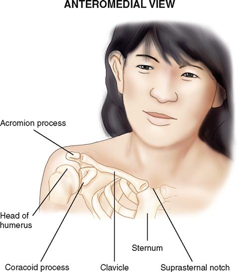
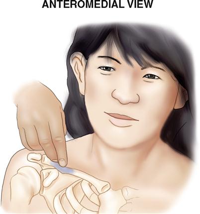
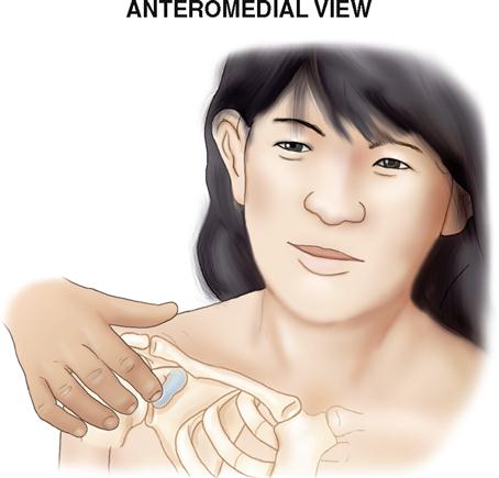
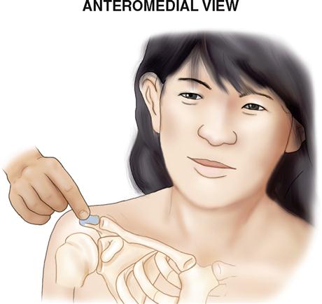
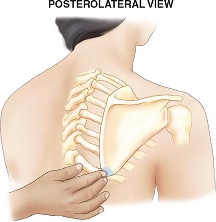
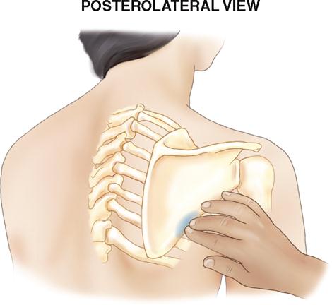
UPPER EXTREMITY
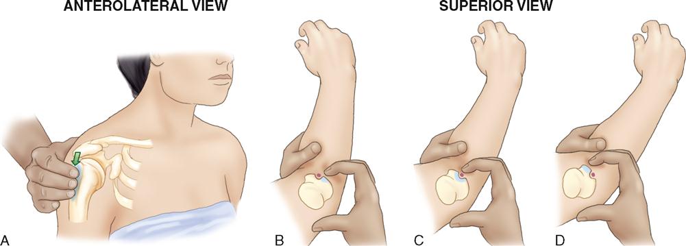
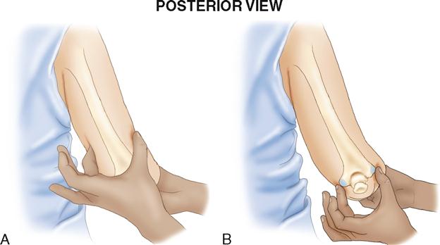
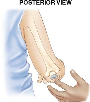
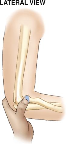
NOTES: (1) If the client’s elbow joint is flexed, the olecranon process will be located farther distally than the two epicondyles of the humerus. (2) Because of the presence of the ulnar nerve, known in lay terms as the “funny bone,” be careful with palpatory pressure between the medial epicondyle of the humerus and the olecranon process of the ulna.
NOTE: A small portion of the distal lateral radial shaft is not directly palpable because it is deep to three deep thumb muscles of the posterior forearm.
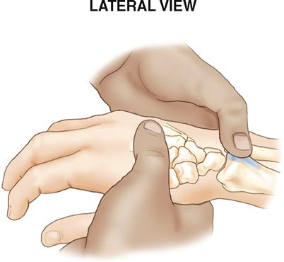
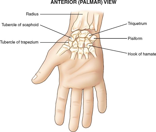
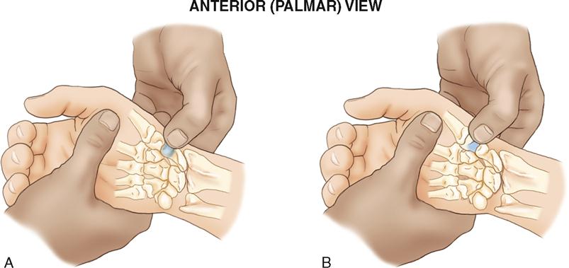
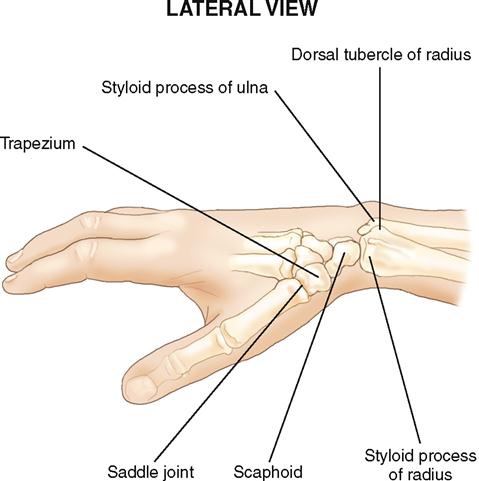
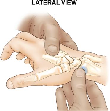
NOTE: The tubercle of the trapezium is located approximately ½ inch distal to the tubercle of the scaphoid.
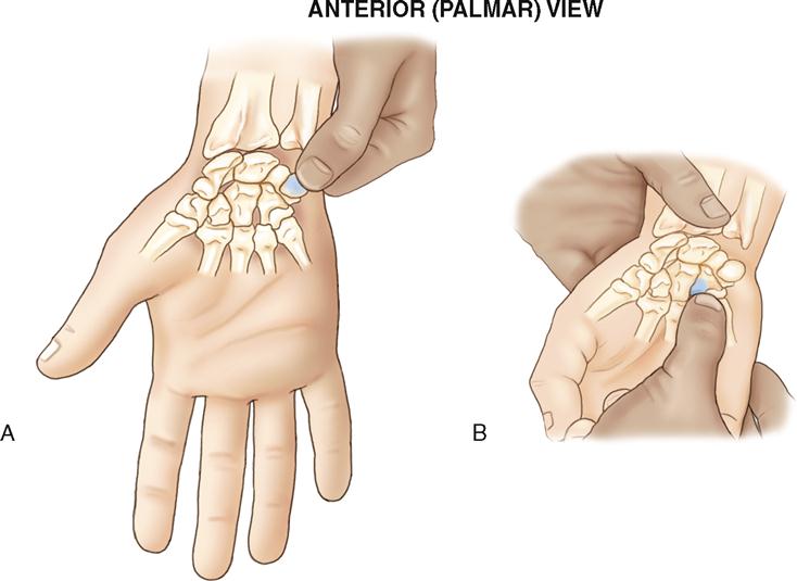
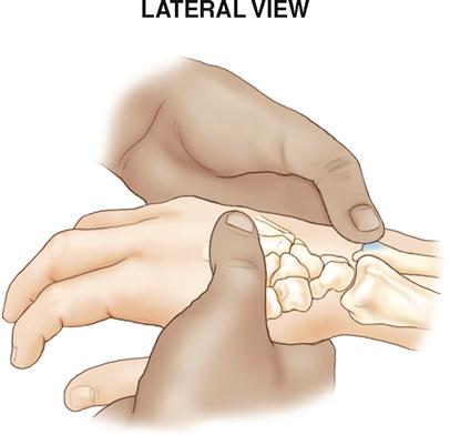
NOTE: The hook of the hamate is fairly pointy and can be somewhat tender to palpation.
AXIAL BODY
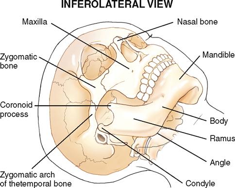
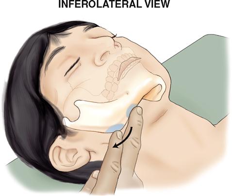
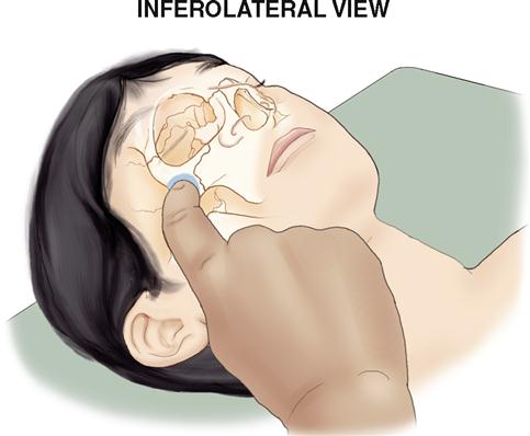
NOTE: The condyle can also be palpated from within the ear. Wearing a finger cot or glove, gently place your palpating finger inside the client’s ear, press anteromedially, and ask the client to alternately open and close the mouth. The movement of the condyle of the mandible at the TMJ will be clearly palpable.
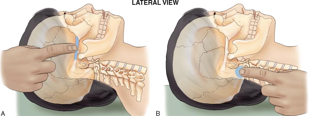
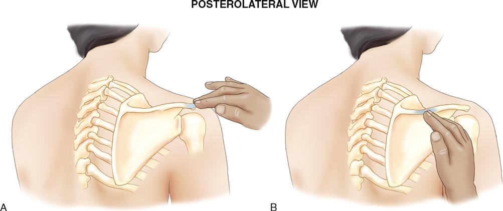
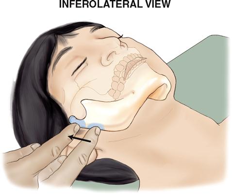
To palpate the mastoid process of the temporal bone, palpate just posterior to the earlobe, then press medially and strum over the mastoid process by moving your palpating finger anteriorly and posteriorly (B).


Stay updated, free articles. Join our Telegram channel

Full access? Get Clinical Tree


