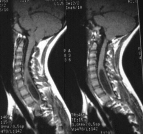
Pitfalls • All cavus foot deformities should be thoroughly worked up for a possible cause for the condition. If the cavus cause is neurologic (Fig. 2), after correcting the bony deformity, later follow-up with tendon transfer to the forefoot or toes is often needed. • This technique is easier and more reliable than the Coleman block test. It allows the surgeon to tell if a sliding calcaneal osteotomy will be necessary to correct heel varus. • The patient’s forefoot is hinded to check the calcaneus position relative to the tibia; if valgus, the forefoot only is corrected. • Using fluoroscopy, needles are placed to identify the midportion of the cuneiform and a location at least 1 cm distal to the physis of the first metatarsal (Figs. 6 and 7).
Osteotomies of the Foot for Cavus Deformities
Indications
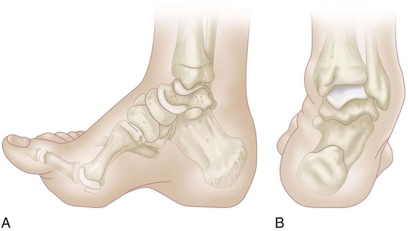
 Rigid cavus foot with plantar flexion of the first ray (Fig. 1A and 1B)
 Ankle/foot instability symptoms, including pain
 Painful metatarsal heads and callosities
Examination/Imaging
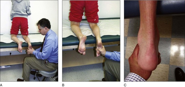
 All patients are assessed to evaluate the flexibility of the hindfoot varus using the kneeling test (Fig. 3A–C).
 Standing anteroposterior (AP) radiographs of both ankles are obtained.
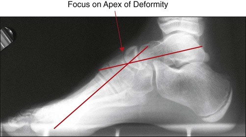
 Standing AP and lateral (Fig. 4) radiographs of both feet are examined, focusing of the apex of the deformity.
Procedure
Step 1: First Ray Osteotomies
 A dorsal closing wedge osteotomy of the first metatarsal and opening wedge osteotomy of the medial cuneiform are performed.
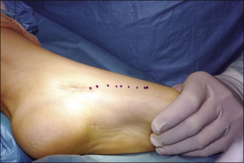
 The first metatarsal and cuneiform are identified through a medial incision on the foot (Fig. 5).
Related posts:
![]() 41: Operative Treatment of Tillaux Fractures of the Ankle
41: Operative Treatment of Tillaux Fractures of the Ankle
 7: Forearm Fractures: Closed Treatment
![]() 32: Patellar Instability: Lateral Release and Medial Plication
32: Patellar Instability: Lateral Release and Medial Plication
![]() 1: Modified Woodward Procedure for Sprengel’s Deformity
1: Modified Woodward Procedure for Sprengel’s Deformity
 40: Proximal Tibial Osteotomy for Blount’s Disease
 60: Thoracoscopic Release and Instrumentation for Scoliosis
![]()
Stay updated, free articles. Join our Telegram channel

Full access? Get Clinical Tree


49: Osteotomies of the Foot for Cavus Deformities


