• 0.5% Marcaine (15–30 ml) is injected: • 0.5% Marcaine (15 ml) is injected in a wheal anteriorly at the level of the ankle joint to anesthetize the branches of the superficial peroneal nerve and sural nerves. • A longitudinal skin incision 4–7 cm long is made dorsomedial and centered over the medial eminence. • Dissection is performed down to the capsule.
Chevron Osteotomy for Adolescent Hallux Valgus
Examination/Imaging
 Anteroposterior, lateral, and oblique weight-bearing films of the foot are obtained and reviewed for hallux valgus.
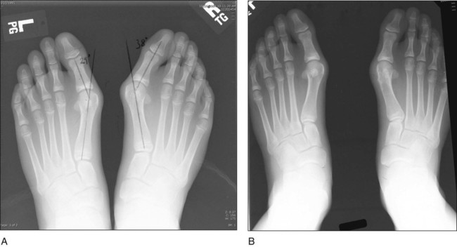
 Figure 1 shows preoperative (Fig. 1A) and postoperative (Fig. 1B) weight-bearing radiographs of a patient treated for hallux valgus.

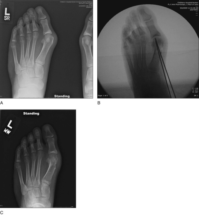
 Figure 2 shows preoperative (Fig. 2A), intraoperative (Fig. 2B), and postoperative (Fig. 2C) radiographs.

Portals/Exposures
 Adjacent to the deep peroneal nerve (near the dorsalis pedis)
 Adjacent to the posterior tibial nerve under the medial malleolus
 Metzenbaum scissors are used to cut through subcutaneous tissue.
 The capsule and metatarsal shaft are exposed with an elevator.
![]()
Stay updated, free articles. Join our Telegram channel

Full access? Get Clinical Tree


48: Chevron Osteotomy for Adolescent Hallux Valgus


