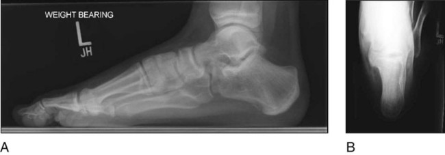
• The patient is observed for an antalgic gait, characterized by a decreased stance phase on the affected extremity, and the position of the foot during stance phase is evaluated. Patients will often present with an out-toeing gait. • Hindfoot and midfoot alignment is examined. Patients with a tarsal coalition will typically present with hindfoot valgus and abduction across the midfoot. • The patient is evaluated for flattening of the medial longitudinal arch. • A single-leg heel rise will assess the flexibility of the flatfoot deformity. Failure to reconstitute the arch of the foot is indicative of a rigid hindfoot and suggests the presence of some abnormality within the subtalar or transverse tarsal joints, such as a tarsal coalition. • Point tenderness may be elicited plantar to the medial malleolus in the region of the sustentaculum tali. • The hindfoot is inverted and everted while stabilizing the ankle joint, and compared to the contralateral side. Tarsal coalitions are associated with pain and limitation of subtalar motion. • Anteroposterior (AP), lateral, and oblique views of the foot are obtained. • The lateral view of the foot (Fig. 1A) is evaluated for a continuous C-shaped line between the posterior aspect of the talar dome and the posterior facet of the subtalar joint (C-sign). • A Harris axial view may demonstrate a middle-facet talocalcaneal coalition (Fig. 1B), although commonly a computed tomography (CT) scan is necessary to adequately identify it. • Standing AP, lateral, and Saltzman hindfoot alignment views may be used to assess alignment. A Saltzman view can be useful to quantify hindfoot valgus.
Resection of Talocalcaneal Tarsal Coalition and Fat Autograft Interposition
Examination/Imaging

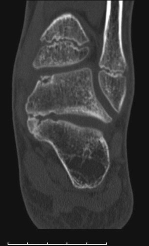
 A CT scan is necessary to better visualize a suspected talocalcaneal coalition, and determine the portion of the subtalar joint that is involved (Fig. 2). The coronal images are the best for examining the subtalar joint.

 Magnetic resonance imaging may be helpful in cases in which the diagnosis is equivocal and a cartilaginous or fibrous coalition is present.
Surgical Anatomy
 Coalitions may form as a result of failure of segmentation of the individual tarsal bones during fetal development.
 Talocalcaneal coalitions occur within the subtalar joint, commonly affecting the middle facet.
 The size of a talocalcaneal coalition is described with respect to the percentage of the subtalar joint that is coalesced.
Portals/Exposures
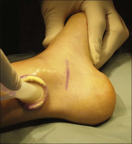
 A horizontal incision is made along the medial aspect of the hindfoot centered over the sustentaculum tali. It should start at the anterior margin of the Achilles tendon and extend distally to the navicular tuberosity (Fig. 3). The skin and subcutaneous tissue are incised sharply.

 The posterior tibial tendon and the flexor digitorum longus (FDL) tendon are identified dorsally. The sheath of the FDL is opened along the length of the incision (Fig. 4A and 4B).
![]()
Stay updated, free articles. Join our Telegram channel

Full access? Get Clinical Tree


46: Resection of Talocalcaneal Tarsal Coalition and Fat Autograft Interposition


