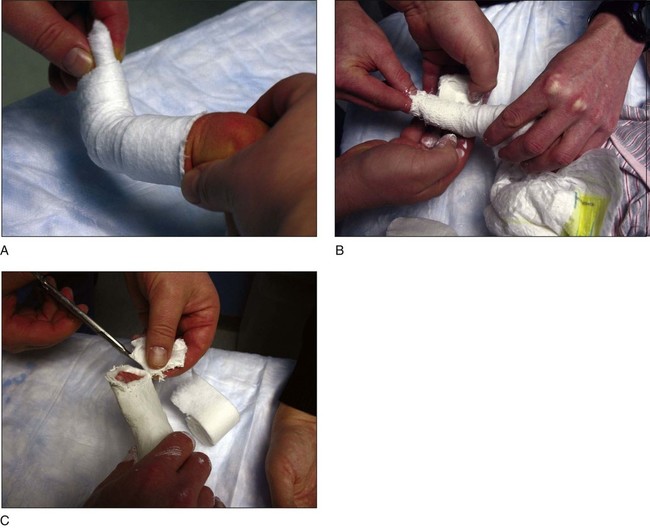
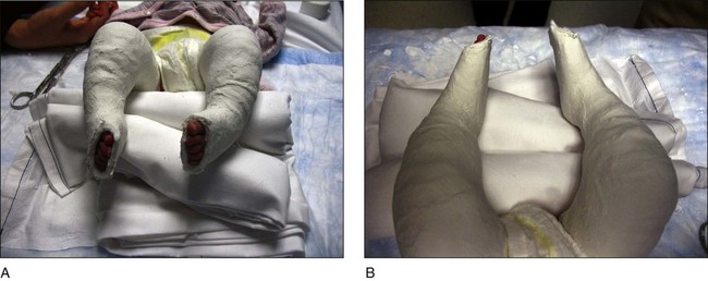
• The forefoot is placed in supination in line with the hindfoot, and the first ray is dorsiflexed. • Cast padding is applied to cover the toes distally (Fig. 3A). A plaster cast is applied using a 2-inch plaster roll (Fig. 3B). Cast should be cut distally so that all five toes are visible (Fig. 3C). • This will correct the cavus deformity. • A short-leg plaster cast is placed first, allowed to dry, and then extended up the thigh to create a long-leg cast. This technique is recommended for all subsequent casts. • Figure 6 shows the first cast for a patient with bilateral clubfoot deformity from below (Fig. 6A) and from above (Fig. 6B).
Ponseti Method for Idiopathic Clubfoot Deformity
Examination/Imaging
 Examination of the foot will demonstrate four components to the deformity:
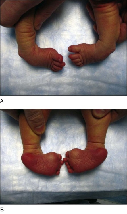
 Figure 1 shows a child with bilateral clubfoot deformity from the dorsal (Fig. 1A) and plantar (Fig. 1B) views.
 The flexibility of the deformity is assessed.
 Deep skin creases are often noted posteriorly and medially.
 There is little role for imaging in the diagnosis of a clubfoot deformity.
Procedure
Step 1
 Manipulations must always be gentle and never painful.
 Approximately 1 minute is spent manipulating the foot.
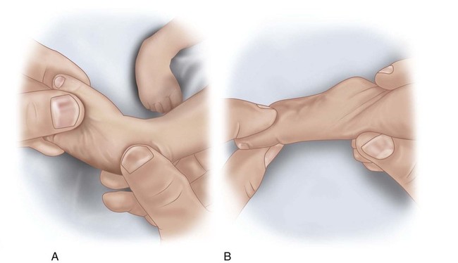
 Figure 2 demonstrates the first manipulation in an anteroposterior (AP) (Fig. 2A) and lateral (Fig. 2B) view. The forefoot is supinated in line with the hindfoot, and the first ray is dorsiflexed.
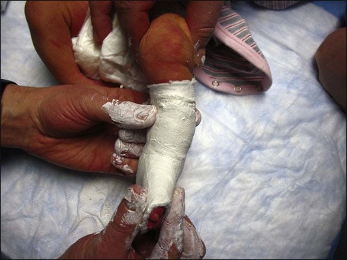
 Correct positioning of the foot is maintained while the short-leg cast is allowed to dry (Fig. 4).
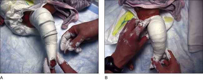
 One the short-leg plaster cast has hardened, the cast is extended up the thigh. Cast padding is applied first (Fig. 5A). Plaster is applied up to the proximal thigh (Fig. 5B).
Step 2
 With the cavus deformity corrected, the next few casts are placed so that the foot is gradually abducted around the head of the talus.
 The foot is manipulated prior to placement of the second cast.
Related posts:
 4: Open Reduction and Internal Fixation of Displaced Medial Epicondyle Fracture Using a Screw and Washer
29: Epiphysiodesis of the Distal Femur/Proximal Tibia-Fibula
32: Patellar Instability: Lateral Release and Medial Plication
31: Discoid Lateral Meniscus
39: Open Reduction and Internal Fixation of Tibial Tubercle Fractures
59: Posterior Instrumented Reduction and Fusion for Spondylolisthesis
4: Open Reduction and Internal Fixation of Displaced Medial Epicondyle Fracture Using a Screw and Washer
29: Epiphysiodesis of the Distal Femur/Proximal Tibia-Fibula
32: Patellar Instability: Lateral Release and Medial Plication
31: Discoid Lateral Meniscus
39: Open Reduction and Internal Fixation of Tibial Tubercle Fractures
59: Posterior Instrumented Reduction and Fusion for Spondylolisthesis
![]()
Stay updated, free articles. Join our Telegram channel

Full access? Get Clinical Tree


