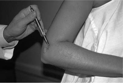
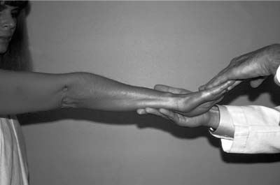
Case 43 A 45-year-old healthy, right-hand–dominant carpenter presents with a 3-month history of activity-related lateral elbow pain. This patient demonstrates normal range of motion of the elbow including normal supination and pronation along with normal stability to valgus, varus, and rotatory stress testing. He is neurovascularly intact and has pain limited to an area immediately distal and anterior to the lateral epicondyle (Fig. 43–1). He also exhibits pain in this same area when wrist extension is resisted by the examiner while the elbow is fully extended (Fig. 43–2). He has no appreciable weakness on examination of his extensor muscles of the forearm. Figure 43–1. The point of maximal tenderness as demonstrated here is almost always just distal and anterior to the lateral epicondyle. Figure 43–2. The examiner is resisting active wrist extension by the patient, generating pain in the area of the lateral epicondyle. • Excise all pathologic tissue in the area of the extensor tendon identified at surgery. • Identifying the area of maximum tenderness in the preoperative holding area helps to localize the site of pathologic tissue. PITFALLS • Take extreme care to avoid the lateral ulnar collateral ligament complex, as transection of the ligament can lead to postoperative posterolateral rotatory elbow instability. • Carefully evaluate the location of the lateral elbow pain to avoid overlooking posterior interosseous nerve compression in the area of the proximal forearm. CONTROVERSIAL POINTS • Arthroscopic tennis elbow release has become more commonly performed. • Some investigators feel that percutaneous release achieves success rates as high as the more extensive standard open release. 1. Radiocapitellar arthritis 2. Radial tunnel syndrome 3. Symptomatic posterolateral plica 4. Meniscoid lesion 5. Lateral epicondylitis Anteroposterior (AP) and lateral radiographs of the elbow fail to reveal any obvious abnormalities. Lateral Epicondylitis.
History and Physical Examination
Differential Diagnosis
Radiologic Findings
Diagnosis
![]()
Stay updated, free articles. Join our Telegram channel

Full access? Get Clinical Tree


