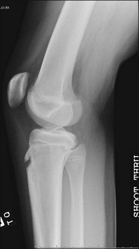
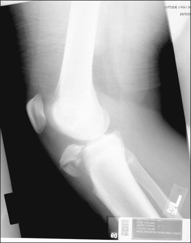
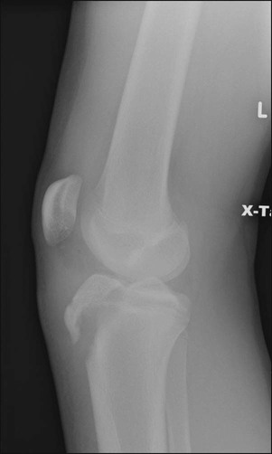
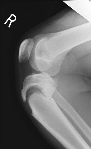
• Figure 2 shows a lateral radiograph of a type I fracture. • Figure 3 shows a lateral radiograph of a type II/III with extension into the joint. • Figure 4 shows a lateral radiograph of a type III fracture hinged at the joint line. • Figure 5 shows a lateral radiograph of a type IV fracture. • When injured, this results in varying degrees of soft tissue disruption to the periosteum, extensor retinaculum, and deep fascia adjacent to the tibial tubercle, which often necessitates repair. Pearls
Open Reduction and Internal Fixation of Tibial Tubercle Fractures
Examination/Imaging
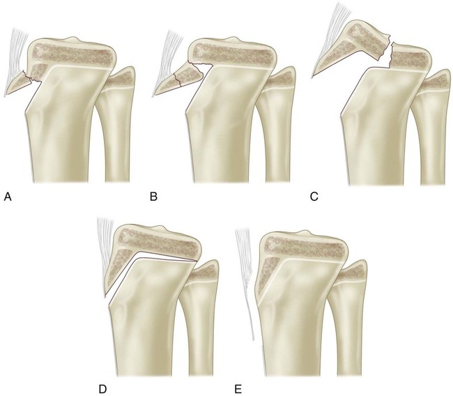
 Fractures can be described by the Ogden modification of the Watson-Jones classification to emphasize both degree of proximal fracture extension and amount of comminution. Figure 1A–E illustrates types I through V, respectively.
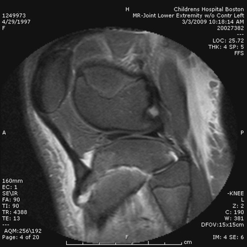
 Magnetic resonance imaging of the knee can show the presence of a nondisplaced fracture line extending into the proximal tibial epiphysis (Fig. 6).
Surgical Anatomy
 The patellar ligament inserts on the tibial tubercle and has attachments into the deep fascia of the proximal tibia.
 Along the lateral aspect of the tibial tubercle, there are branches of the anterior tibial recurrent artery that may retract laterally and distally into the musculature of the anterior compartment at the time of injury, resulting in hematoma formation and increasing the risk of developing compartment syndrome (Fig. 7).
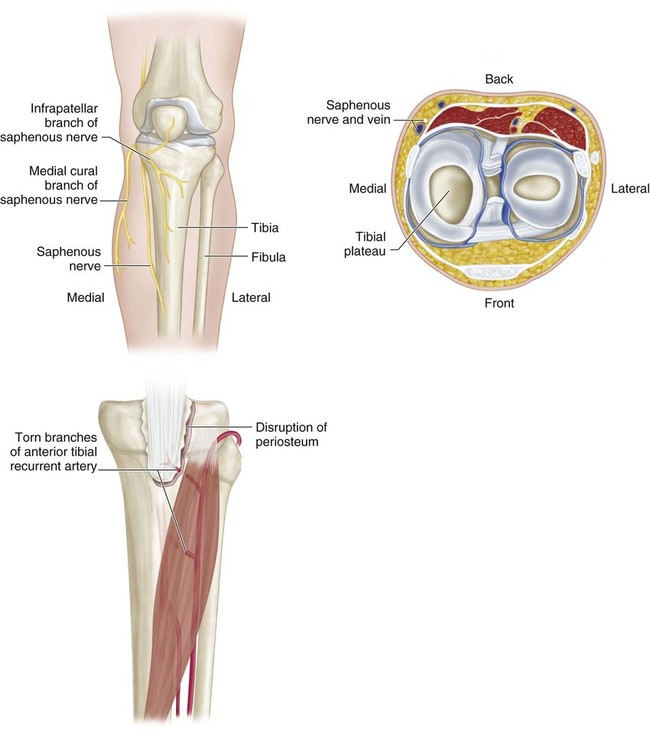
 Medial branches of the infrapatellar branch of the saphenous nerve may be observed, and care should be taken to preserve this nerve to avoid a large area of anesthesia.
Positioning
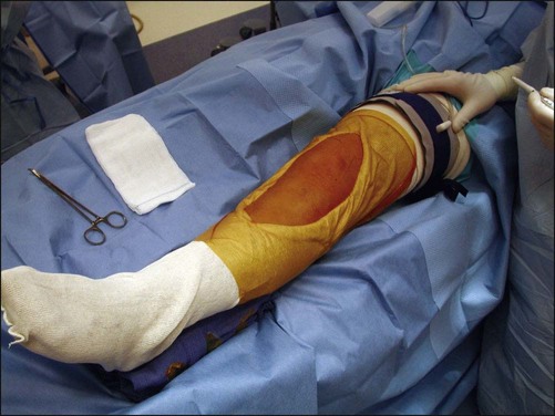
 The patient is positioned supine on a radiolucent table with a padded sterile tourniquet placed around the proximal thigh (Fig. 8).
Related posts:
![]() 41: Operative Treatment of Tillaux Fractures of the Ankle
41: Operative Treatment of Tillaux Fractures of the Ankle
 7: Forearm Fractures: Closed Treatment
![]() 32: Patellar Instability: Lateral Release and Medial Plication
32: Patellar Instability: Lateral Release and Medial Plication
![]() 1: Modified Woodward Procedure for Sprengel’s Deformity
1: Modified Woodward Procedure for Sprengel’s Deformity
![]() 47: Resection of Calcaneonavicular Coalition and Fat Autograft Interposition
47: Resection of Calcaneonavicular Coalition and Fat Autograft Interposition
 60: Thoracoscopic Release and Instrumentation for Scoliosis
![]()
Stay updated, free articles. Join our Telegram channel

Full access? Get Clinical Tree


39: Open Reduction and Internal Fixation of Tibial Tubercle Fractures


