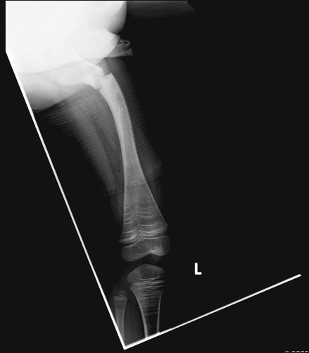
Sofield Osteotomy with Intramedullary Rod Fixation of the Long Bones of the Femur/Tibia
Positioning
 The patient is placed supine on a radiolucent table.
 Intraoperative fluorscopy is utilized.
 The entire gluteal area must be draped free to access the greater trochanter for rodding; the entire lower extremity is prepped out.
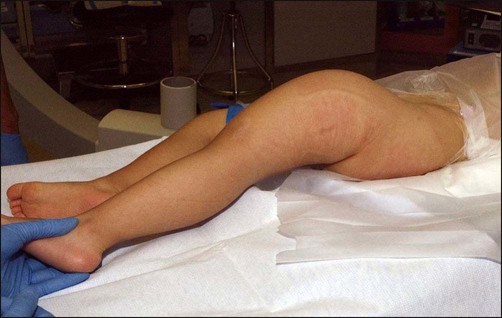
 For surgery on one lower extremity, a trochanter roll is appropriate (Fig. 2), with the ipsilateral arm across the chest.
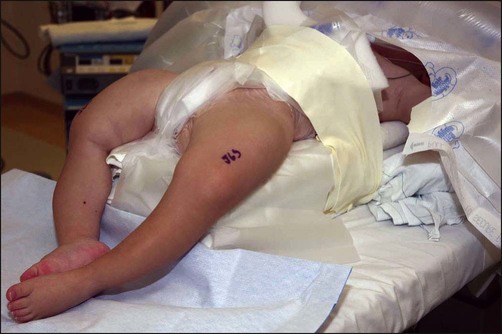
 For surgery on bilateral lower extremities, access to both gluteal areas is ensured using a stack of blankets to elevate the patient if needed (Fig. 3).
Portals/Exposures
Step 1 (Approach A)
 The entry point to the greater trochanter is identified. Figure 4 shows the trochanteric entry point for a femoral rodding with a Schnidt forceps marking the first osteotomy.
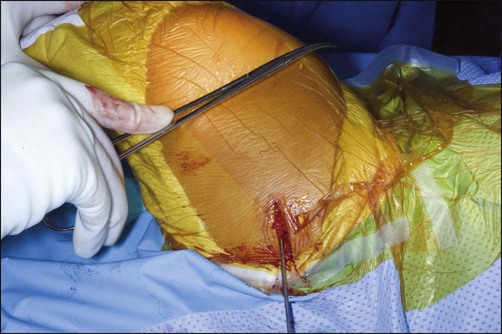
 The incision needs to be posterior and inferior to the tip of the greater trochanter. A 2-cm incision is sufficient.
 Dissection is performed bluntly and longitudinally, spreading through the gluteus maximus until the tip of the greater trochanter can be accessed.
Procedure
Step 1 (Approach B)
 The fracture site is accessed first, and a guidewire is passed retrograde through the greater trochanter (Fig. 5). The guidewire is drilled into the greater trochanter and down the canal, and AP and lateral radiographs are checked for position.
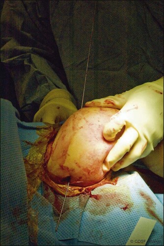
 Drilling stops when the wire is at a cortex; this will be the apex of the deformity and the appropriate position for the first osteotomy.
![]()
Stay updated, free articles. Join our Telegram channel

Full access? Get Clinical Tree


30: Sofield Osteotomy with Intramedullary Rod Fixation of the Long Bones of the Femur/Tibia


