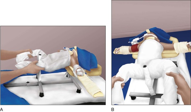
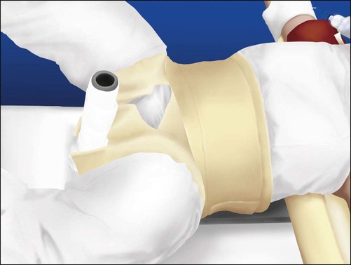
• Proximal-third fractures can be hard to maintain. The proximal fragment tends to be abducted and flexed by the insertion of the muscles at the trochanters. • Middle-third fractures tend to fall into varus and external rotation. A valgus mold is important to resist this deformity. • For fractures of the distal third of the femur, the gastrocnemius tends to pull the fracture into recurvatum. Flexing the knee takes tension off the muscle and helps to maintain the reduction.
Femur Fracture
Closed Reduction and Spica Cast
Anatomy
 In order to perform the appropriate reduction and mold the cast, it is important to remember the muscles forces acting on the fracture.


Related posts:
 7: Forearm Fractures: Closed Treatment
 60: Thoracoscopic Release and Instrumentation for Scoliosis
Stay updated, free articles. Join our Telegram channel

Full access? Get Clinical Tree


