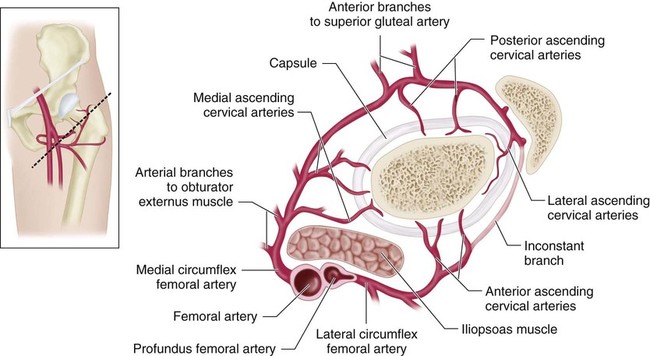
Femur Fracture
Lateral Trochanteric Entry Rigid Nailing
Indications
 Rigid intramedullary (IM) nail fixation for femoral diaphyseal fractures in children and adolescents over the age of 8 years until skeletal maturity.
 All fracture patterns, including transverse, spiral, oblique, and comminuted fractures, can be successfully managed with this technique.
 This technique is particularly useful in unstable fracture patterns.
 The technique can also be used for stabilization of femoral diaphyseal osteotomies in patients age 8 years to skeletal maturity.
Examination/Imaging
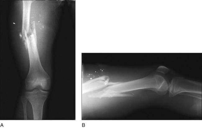
 Standard anteroposterior (AP) (Fig. 1A) and lateral (Fig. 1B) radiographs of the femur as well as of the ipsilateral hip and knee should be obtained.
 In patients with comminuted fractures, radiographs of the contralateral femur can be used to determine the length of the injured femur.
Positioning
 The patient may be positioned in the hemilithotomy position on a fracture table. Traction is applied through a boot (Fig. 3). The post should be well padded where it contacts the perineum.
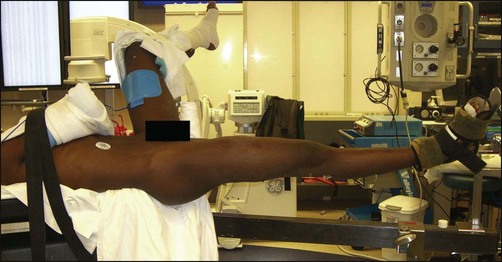
 As an alternative, the patient may be positioned supine on a radiolucent table. This is the preferred table for performing osteotomies. If this is used for fractures, an assistant will be needed to apply traction to the lower extremity to bring the fracture out to length.
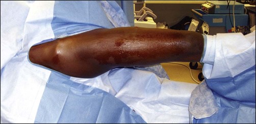
 An image intensifier should be draped for visualization of the femur. Figure 4 shows draping of the extremity.
Portals/Exposures
 The image intensifier is used to assess the entry point. Because the proximal fragment tends to externally rotate on the fracture table, it is helpful to arc the image intensifier over the patient’s hip to visualize the lateral aspect of the greater trochanter.
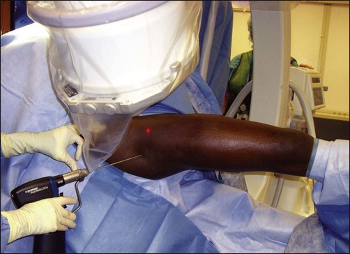
 A 3.2-mm guidewire is placed via percutaneous entry (Fig. 5). Using fluoroscopic guidance, the tip of the guidewire is placed into the lateral aspect of the greater trochanter.
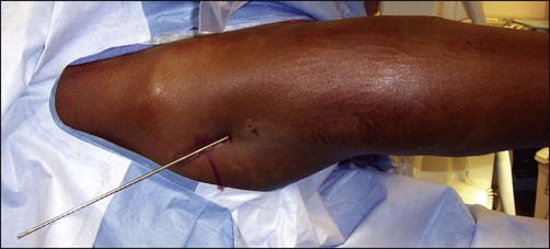
 The guidewire is advanced into the medullary canal aimed slightly medially.
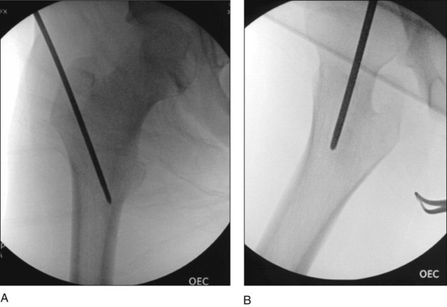
 Placement of the guidewire is confirmed by fluoroscopy in both the AP (Fig. 6A) and lateral (Fig. 6B) planes.
 A small incision (approximately 1.5 cm) is made extending proximal from the insertion site of the guidewire to allow for passage of the entry reamer.
Musculoskeletal Key
Fastest Musculoskeletal Insight Engine


