Pearls
Greater Trochanteric Transfer/Relative Femoral Neck Lengthening
Surgical Anatomy
 Fascial interval between tensor and gluteus maximus
 Greater trochanter and abductor insertion on trochanter
 Piriformis tendon and short external rotator tendons
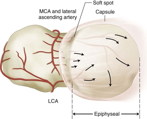
 Medial femoral circumflex artery (MFCA) along its course posterior to greater trochanter, from the level of quadratus femoris until its terminal branches into the posterolateral/lateral aspect of the femoral head (Fig. 4)
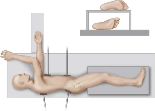
Portals/Exposures
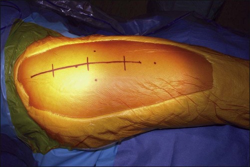
 A direct lateral skin incision is made; fasciotomy by the Gibson approach is preferred. The incision is continued in the proximal interval between the posterior edge of the tensor fasciae latae (TFL) and the anterior edge of the gluteus maximus (Fig. 6).
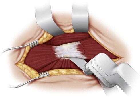
 The plane for the posteroanterior osteotomy of the greater trochanter is at a level just superficial to the piriformis tendon insertion onto the posterosuperior portion of the base of the trochanter (Fig. 7).
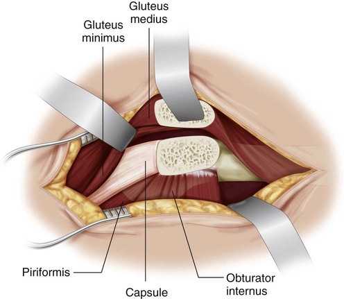
 The proximal plane lies between the posterolateral capsule and the tip of the greater trochanter (this dissection is most safely done subperiosteally, hugging the bone of the posterosuperior edge of the greater trochanter) (Fig. 8).
Related posts:
![]() 41: Operative Treatment of Tillaux Fractures of the Ankle
41: Operative Treatment of Tillaux Fractures of the Ankle
 7: Forearm Fractures: Closed Treatment
![]() 32: Patellar Instability: Lateral Release and Medial Plication
32: Patellar Instability: Lateral Release and Medial Plication
![]() 18: Percutaneous in situ Cannulated Screw Fixation of Slipped Capital Femoral Epiphysis
18: Percutaneous in situ Cannulated Screw Fixation of Slipped Capital Femoral Epiphysis
![]() 47: Resection of Calcaneonavicular Coalition and Fat Autograft Interposition
47: Resection of Calcaneonavicular Coalition and Fat Autograft Interposition
 60: Thoracoscopic Release and Instrumentation for Scoliosis
![]()
Stay updated, free articles. Join our Telegram channel

Full access? Get Clinical Tree


24: Greater Trochanteric Transfer/Relative Femoral Neck Lengthening