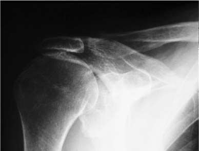
Case 23 A 69-year-old man presents with a 12-month history of shoulder pain and loss of motion. He reports the initiation of symptoms after a fall in his garden. Since that time, his pain and functional losses have slowly but persistently progressed. In the 6 weeks prior to presentation, he has noticed a significantly reduced ability to raise his arm in front of his body. He denies any numbness or other symptoms. Active forward flexion is limited to 40 degrees and passive forward flexion is 160 degrees. Active external rotation is 40 degrees, but only 3/5 external rotation strength is present. He has mildly painful crepitation to passive range of motion of the shoulder and has a negative lift-off test. There is also some atrophy of the supraspinatus and infraspinatus muscles within their respective fossae. The patient is not tender to palpation about the shoulder and otherwise has no abnormalities. Figure 23–1. An anteroposterior (AP) radiograph of the affected shoulder. 1. Suprascapular nerve injury 2. Massive rotator cuff tear 3. Glenohumeral arthritis An anteroposterior view of the shoulder is presented (Fig. 23–1). Massive Rotator Cuff Tear. This patient’s history of a fall that initiated his symptoms of weakness and pain suggests a rotator cuff tear. His progressive loss of function associated with his physical findings of crepitation and supraspinatus and infra-spinatus atrophy suggests a very large to massive tear. Although his physical findings and symptoms could be due to a suprascapular nerve injury associated with his original fall, such an injury is very uncommon. Sophisticated imaging studies such as magnetic resonance imaging could help confirm rotator cuff tearing, but this patient’s physical examination and history are highly suggestive. Important keys to differentiating smaller tears from massive tears include the poor active forward flexion that this patient exhibits as well as the muscle atrophy and very weak external rotation that is seen clinically. The negative lift-off test confirms the intact nature of the subscapularis tendon. An organized rotator cuff program is recommended for almost all patients presenting with symptoms suggestive of rotator cuff tearing. Even with massive rotator cuff tears, nonsteroidal antiinflammatory medications and organized rotator cuff strengthening can often improve function significantly. An important consideration regarding these functional gains relates to the age, activity level, and goals of an individual patient. Failure of therapy and other nonoperative measures to improve function and motion adequately is an indication for surgical intervention. Complete repair of massive rotator cuff repairs is sometimes impossible, making intraoperative decisions regarding the viability of partial rotator cuff repair or simple debridement important. Several methods for determining rotator cuff size have been described, but the authors prefer to describe rotator cuff tears by the area of the defect. Using this classification system, massive rotator cuff tears are defined as those tears occupying an area of 20 cm2. As an example, this represents a defect of 5 4 cm, or 20 cm2. Although characterization of the size and dimensions of the tear is very important in helping to determine the viability of complete rotator cuff repair, the size of the tear should not be used as the only criterion to determine treatment. Often, massive rotator cuff tears can be mobilized to a great extent so as to allow for complete repair. Conversely, some smaller rotator cuff tears can be mobilized very little, making complete repair impossible. For this reason, intraoperative assessment of the quality and mobility of the tendons is one of the most important determinations that the surgeon must make. Patients failing to adequately respond to a nonoperative treatment program are candidates for surgical intervention. A number of alternatives for treatment of massive rotator cuff tears have been described in the literature. These range from extensive muscle slide operations, the supplementation of graft materials in an attempt to repair the cuff, as well as operations designed only to debride rotator cuff tissue. However, no procedure is the best operation for all patients with massive rotator cuff tears. The authors feel that each case must be individualized, based on the quality and mobility of the rotator cuff tissue as determined at the time of surgery. The authors perform shoulder arthroscopy initially on all patients with rotator cuff tears. There are several advantages to arthroscopic evaluation. First, the arthro-scope allows for a complete assessment of the glenohumeral joint, and an accurate evaluation of the size of the rotator cuff tendon tear and of the status of the biceps tendon, as well as an opportunity to debride these tissues. Second, the arthroscope can be used to determine both the quality and the mobility of the residual rotator cuff tendon. Small graspers and arthroscopic freers will allow for an evaluation of the mobility of the rotator cuff tendon and thus an assessment of the reparability of the tear. The authors have found that the large majority of massive rotator cuff tears respond to a partial repair when a complete repair is not possible. The concept of a partial rotator cuff repair as promoted by Burkhart and colleagues (1998) has proved very valuable to the authors in the management of these complex and extensive rotator cuff tears. The technique of partial repair involves repair of the margins of the tear to restore the force couples and “suspension bridge” system of force transmission in the shoulder. Complete coverage of the defect is not considered essential as long as the normal mechanics of the shoulder are restored and the rotator cuff tear is converted to a functional rotator cuff tear. The procedure begins with the patient either in the beach chair or lateral decubitus position. Standard anterior and posterior arthroscopic portals are established and glenohumeral joint visualization accomplished. Care is taken to assess the articular surfaces of both the glenoid and humeral head, as significant degeneration is not uncommonly seen in patients with massive rotator cuff tears. Glenohumeral joint debridement is carried out with attention paid to the axillary pouch where loose bodies may occasionally be found. The status of the biceps tendon is also evaluated, as biceps tendon ruptures or near-complete ruptures are commonly seen in this clinical situation. Following debridement, attention is turned to the subacromial space where debridement allows for visualization of the margins of the rotator cuff tear. It is important to carefully assess the quality of the tissue seen in and around the rotator cuff, as thickened bursal tissue may often look much like rotator cuff tendinous tissue. Following debridement of this tissue and identification of the rotator cuff residual rim, the size of the tear can be accurately determined. Arthroscopic instruments of known diameter can be used to make this determination. Arthroscopic graspers and arthroscopically placed sutures can be used to apply both lateral traction to the supraspinatus tendon and/or anterior traction to the more posteriorly located infraspinatus and teres minor musculotendinous units. The subscapularis tendon must also be carefully visualized particularly when in the glenohumeral joint because partial or even complete subscapularis tendon tears occur as well. Small arthroscopic freers and arthroscopic shavers can be used to help mobilize the residual rotator cuff tendinous tissue. Following mobilization, an assessment can be made as to the reparability of the massive rotator cuff tendon tear. Often it is apparent that a complete repair of the residual tissue would be impossible, but careful evaluation will allow the surgeon to recognize that reapproximation of a large portion of the infraspinatus tendon will help to balance this “dysfunctional” rotator cuff tear and restore the force couples. If a partial rotator cuff repair is to be attempted, the authors first perform an arthroscopic acromioplasty using standard technique. When preoperative acromioclavicular joint symptoms are present, an arthroscopic distal clavicle excision is performed as well. The authors perform the great majority of partial and complete rotator cuff repairs through arthroscopic technique. Arthroscopic repair of massive tears is technically challenging but possible using a systematic approach. Extensive experience in the repair of smaller rotator cuff tears is essential before the surgeon attempts repair of more massive tears. Often, extensive mobilization of the tendon is required, and a combination of convergence su tures and anchor fixation is necessary. Preparation for the arthroscopic repair, including decompression and distal clavicle resection with humeral head preparation and cuff mobilization must be accomplished relatively quickly since these patients often tend to have increased soft tissue swelling as a consequence of their general tissue quality. Also, the absence of rotator cuff tissue or the inability to adequately mobilize the cuff back to the articular rim of the humerus may necessitate a partial rotator cuff repair in some of these patients. However, it is much better to perform a balanced partial repair of the available cuff tissue that will function well than to perform a complete repair that is under high tension postoperatively. Identification of the rotator cuff configuration and orientation is critical in maximizing the repairability of these massive rotator cuff tears. An examination under anesthesia is important to perform at the initiation of these procedures. Capsular contracture causing loss of passive motion can often be restored with a gentle manipulation making rotator cuff repair more easily performed since the tendency of the humeral head to sublux will be lessened by the inferior capsular release. Both the beach chair position and lateral decubitus position are acceptable for massive rotator cuff repairs. Gentle traction on the arm in the beach chair position often aids not only in visualization of the subacromial space, but also since it allows for more effective lateralization of the cuff in many instances. Standard arthroscopy portals are usually all that is required, although accessory portals may aid in accessing certain portions of the cuff tear or in allowing for traction sutures to be utilized more effectively during cuff mobilization. The articular examination is very important in these patients with massive cuff tears. An assessment of the biceps and articular surfaces can be carried out. Often, significant articular wear or degeneration is present in these patients, and this may have implications both in the postoperative rehabilitation program and as it pertains to the chances for a successful postoperative outcome. Synovitis is also often present within the glenohumeral joint, and a partial synovectomy can be performed. Finally, if a gentle manipulation is not successful and significant passive motion is still present, an arthroscopic capsular release can significantly improve mobility of the humeral head to allow for a more concentric reduction onto the glenoid in some instances. Arthroscopic subacromial bursa evaluation allows for the excision of bursal tissue and the opportunity to define the scale and extent of the rotator cuff tear. Use of a grasping device can aid significantly in determining the extent of mobilization required and the orientation of the rotator cuff tendon tear itself. Not uncommonly, massive tears may be very easily lateralized back to the tuberosities, and minimal mobilization is required. This subacromial arthroscopy also allows for an assessment of the reparability of the cuff tear itself. An assessment of rotator cuff tendon quality as well as the shape of the tear and mobility of the tear all play a role in this determination. Once the determination that a significant partial repair or complete repair can be accomplished, arthroscopic subacromial decompression often combined with distal clavicle resection is carried out. The coracoacromial arch is preserved only in those cases where little functional cuff tissue can be effectively reap-proximated to the tuberosities. The tuberosities can also be prepared by debridement of the soft tissue and very limited abrasion of the tuberosities themselves. This bone is often very osteopenic, and anchors are generally more likely to not achieve adequate fixation than in smaller tears. The first step in mobilizing a massive cuff tear is often to release the superior capsule from above the glenoid. This is accomplished by first removing any reactive synovium present in the superior gutter. The capsule can then be released with the arthroscopic shavers or with a punch. PEARLS • The arthroscope is an extremely valuable tool in the assessment of massive rotator cuff tears. It allows for an evaluation of the amount, quality, and mobility of the residual tendinous tissue. • When debridement and acromioplasty are accomplished arthroscopically, the mini-open deltoid splitting approach, when necessary, almost always allows for excellent repair of the rotator cuff tear. The components of the tear can be brought into the field of view through the limited arthrotomy by having an assistant simply rotate the arm to accommodate for visualization and repair.
History and Physical Examination
Differential Diagnosis
Radiologic Findings
Diagnosis
Surgical Management
Related posts:

Stay updated, free articles. Join our Telegram channel

Full access? Get Clinical Tree


