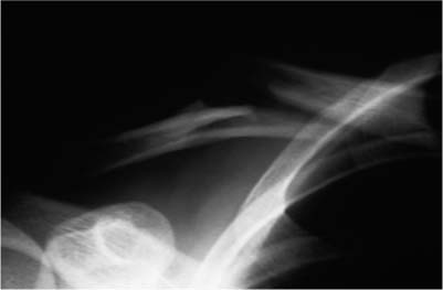
Case 10 A 26-year-old recreational athlete presents 2 days after a fall onto the right shoulder while playing touch football. He complains of pain and swelling in the area of the clavicle and denies any numbness or weakness in the arm. He also denies any neck injury or other abnormality. Moderate swelling and tenderness is present in the area of the midclavicle. Passive range of motion of the shoulder is normal, although motion causes moderate pain and some crepitus in the area of the midclavicle. The sternoclavicular joint is mildly tender, as is the acromioclavicular joint, but no obvious deformity of either joint is seen. Careful neurovascular examination fails to demonstrate any abnormalities. The patient’s neck is supple with a full range of motion, and his breath sounds are symmetric. Figure 10–1. An anteroposterior (AP) radiograph of the clavicle and shoulder. 1. Pectoralis major muscle rupture 2. Midshaft clavicle fracture 3. Subcutaneous emphysema 4. Rotator cuff tear An anteroposterior (AP) radiograph is shown (Fig. 10–1). Midshaft Clavicle Fracture. Tenderness, swelling, and crepitus in the area of the clavicle are all suggestive of an acute clavicle fracture. While tenderness is present in the area of the pectoralis muscle and clavicle, shoulder range of motion is good. Radiographs will almost always identify middle third clavicle fractures, but displace ment in the axial plane is sometimes difficult to identify on AP X-rays alone. Computed tomography (CT) scans or apical oblique radiographs may aid in assessing displacement in this plane. A careful neurovascular examination is important in all clavicle fractures, as the proximity of the neurovascular structures increases their risk of injury. PEARLS • Most middle third clavicle fractures can be treated symptomatically. The use of an arm sling and/or figure-of-eight harness usually provides good symptomatic relief in the first week or two. However, the authors rarely require immobilization and make an effort to explain that the figure-of-eight harness is used purely for symptomatic relief and that it is not felt to be necessary or helpful in increasing the chances of fracture union or improving fracture alignment. • General shoulder motion exercises including pendulums and elbow motion exercises are important in maintaining joint function in the first few weeks following a clavicle fracture. • Smokers are encouraged to stop smoking during the healing phase to encourage clavicle union. PITFALLS • Inadequate internal fixation increases the risk of nonunion significantly. A rigid plate and screw construct provides maximum stability when open reduction and internal fixation of midshaft clavicle fractures is required. • The great majority of midshaft clavicle fractures heal with nonoperative treatment. The indications for operative fracture treatment should remain limited to those clinical situations where closed treatment is deemed unreasonable. Clavicle fractures are grouped according to their position on the clavicle. Middle third clavicle fractures are considered group I fractures. Group II fractures constitute fractures of the distal third, and these fractures can be further subtyped according to their position in the distal third of the shaft. Group III fractures constitute fractures of the proximal one third of the clavicle. Group I fractures, or fracture of the middle third, are the most common fractures seen in adults and children. They generally occur at a point in which the clavicle changes to a flattened cross section. This area of the clavicle is otherwise unsecured, as opposed to the more proximal and distal segments, which are mechanically supported by the ligamentous structures and muscular attachments at each end. Middle third (group I) fractures account for approximately 80% of all clavicle fractures. Group II fractures account for 15% of all clavicle fractures and are subtyped according to their location relative to the coracoclavicular ligaments and the articular surface of the distal clavicle. Neer emphasized the importance of subdividing these Group II fractures according to their location and articular extension. Type I fractures of the distal third occur lateral to the coracoclavicular ligaments but do not extend into the acromioclavicular joint articulation. Because the ligaments remain intact, there is little displacement of the fracture fragments. The great majority of midshaft clavicle fractures can be treated nonoperatively. In general, excellent results have been reported with nonoperative treatment of these fractures. Most authors who have written on the treatment of these clavicle fractures have recommended a conservative approach. The goal of treatment for clavicle fractures is to achieve bone healing with minimum morbidity, minimal loss of function, and minimal residual deformity.
History and Physical Examination
Differential Diagnosis
Radiologic Findings
Diagnosis
Surgical Management
Related posts:

Stay updated, free articles. Join our Telegram channel

Full access? Get Clinical Tree


