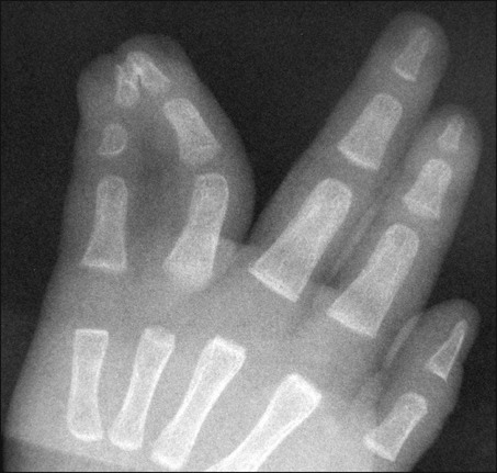
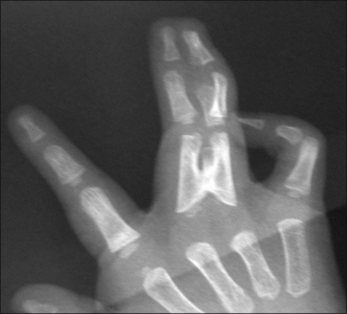
• Most surgeons recommend waiting until the child is at least 6 months old to allow tissues to grow and to reduce the anesthetic risk to the child, and some authors recommend waiting until the child is over the age of 2. All surgical releases should be completed before the child reaches school age. • An exception to waiting until the child is 6 months of age is when the syndactyly involves the ring and small finger or the thumb and index finger. These are released earlier (between 3 and 6 months) due to the risk of developing worsening deformity due to the differential growth rates of these digits (Fig. 1). • Presence or absence of independent motion of the digits. • The presence of joint creases can help determine if the joints are functioning and motion is present. • The digital artery and nerve are often easiest to identify proximally. Proper digital arteries that bifurcate distal to the planned web space will need to be ligated. If this is necessary, the proper digital artery to the more midline digit is usually preserved. • Digital nerves that bifurcate distal to the planned web space can be separated by interfasicular microdissection. • A normal web space is wider distally than proximally and begins its dorsal-to-palmar slope of about 35–45° at the level of the metacarpal head. • Many different local skin flaps have been described to re-create the web space. • Care is taken at the beginning of the procedure to plan the location of the web space such that it will follow the normal cascade of the hand.
Digital Syndactyly Release
Indications
 Surgical separation is indicated in almost all cases as syndactyly can have functional, cosmetic, and developmental implications that can worsen with growth of the child.

 While the most common indication for surgical separation is functional limitations caused by the webbed digits, many digits are also separated for cosmetic reasons.
Examination/Imaging
Physical Examination
 A detailed examination of both upper extremities needs to be performed and, occasionally, a more detailed examination of the child is necessary to rule out any associated syndromes.
 Specific details to note include:
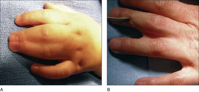
 Complete versus incomplete. Figure 2A shows a complete syndactyly involving the long and ring fingers. Figure 2B shows an incomplete syndactyly involving the same fingers.


 Features that may indicate associated syndromes include:
Surgical Anatomy
 As the digits are separated, the neurovascular structures must be identified and protected.
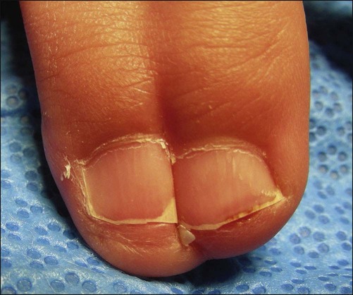
 The lateral eponychial fold is often absent in complex or simple complete syndactyly. It can be reconstructed using tissue from the distal tip of the adjoined digit, and its careful reconstruction will improve the cosmetic outcome of the procedure.
Musculoskeletal Key
Fastest Musculoskeletal Insight Engine


