Muscles of the Leg and Foot
The muscles of this chapter are primarily involved with motions of the foot at the ankle and subtalar joints and/or the motions of the toes at the metatarsophalangeal (MTP) and interphalangeal (IP) joints.
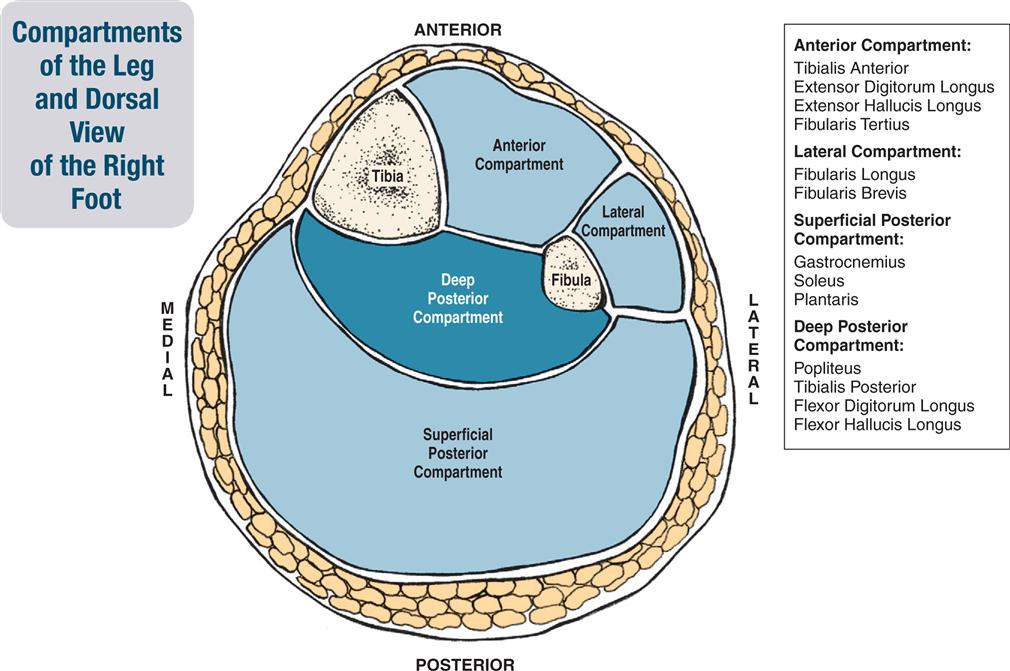
As a rule, muscles that move the foot originate (attach proximally) and have their bellies in the leg. Leg muscles are usually divided into the four fascial compartments of the leg: anterior, lateral, superficial posterior, and deep posterior.
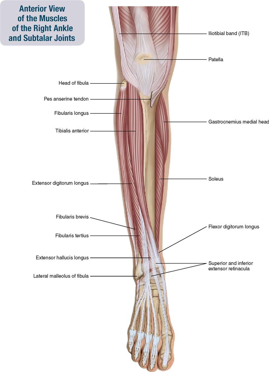
The anterior compartment contains the tibialis anterior, extensor digitorum longus, extensor hallucis longus, and fibularis tertius.
The lateral compartment contains the fibularis longus and fibularis brevis.
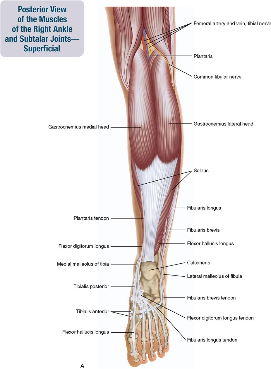
The superficial posterior compartment contains the gastrocnemius, soleus, and plantaris.
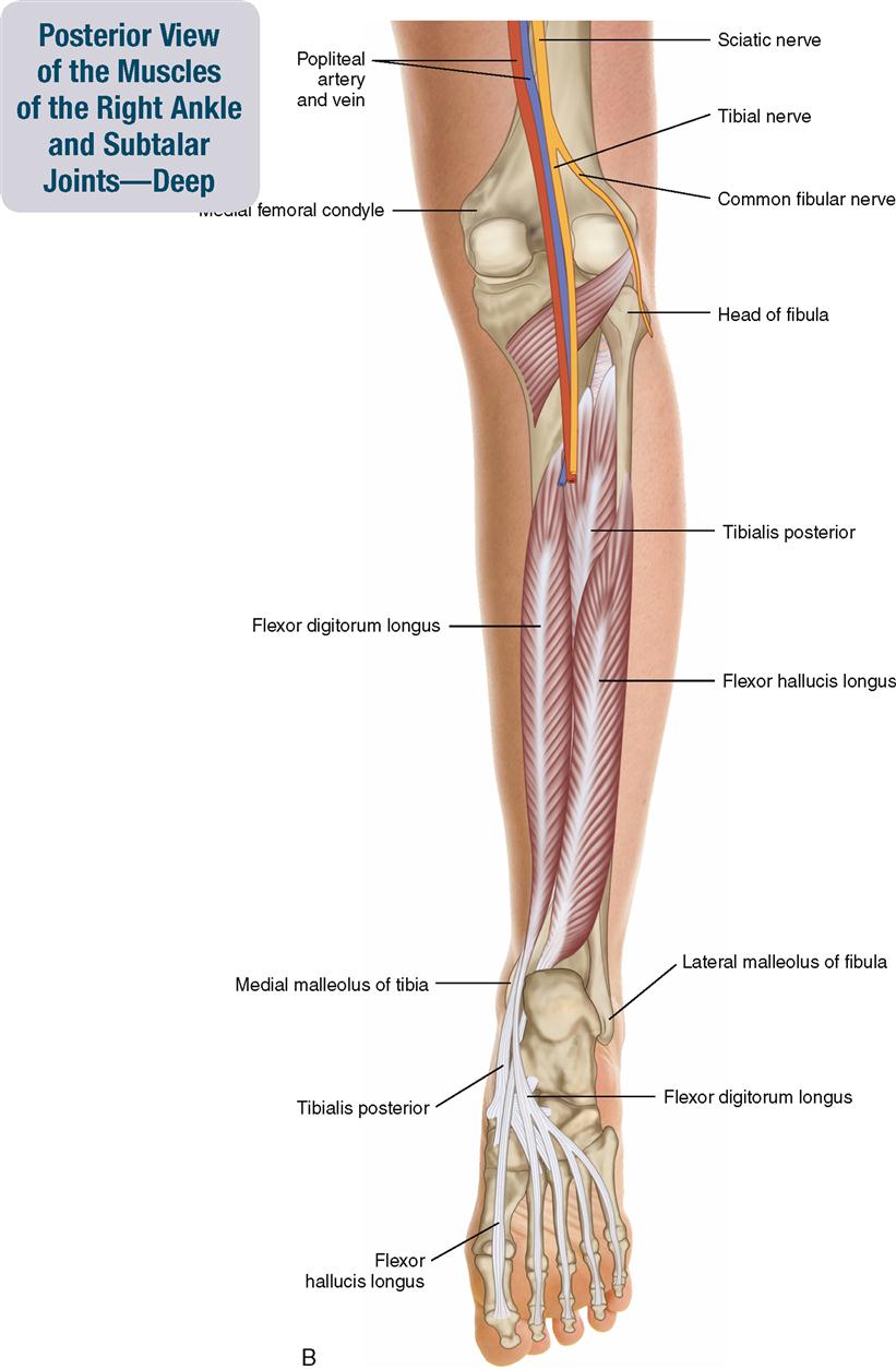
The deep posterior compartment contains the tibialis posterior, flexor digitorum longus, flexor hallucis longus, and popliteus. Some of these muscles are addressed in other chapters.
The location by compartment helps determine the actions of these muscles. For example, all muscles of the anterior compartment perform dorsiflexion; all muscles of the lateral and posterior compartments perform plantarflexion; and all muscles of the lateral compartment perform eversion. The final determination of exactly what the actions of a muscle of the ankle and subtalar joints will be is where the distal tendon of that muscle crosses these joints (e.g., the belly of the tibialis anterior is located in the anterolateral leg, but its tendon crosses the subtalar joint medially; therefore it inverts the foot).
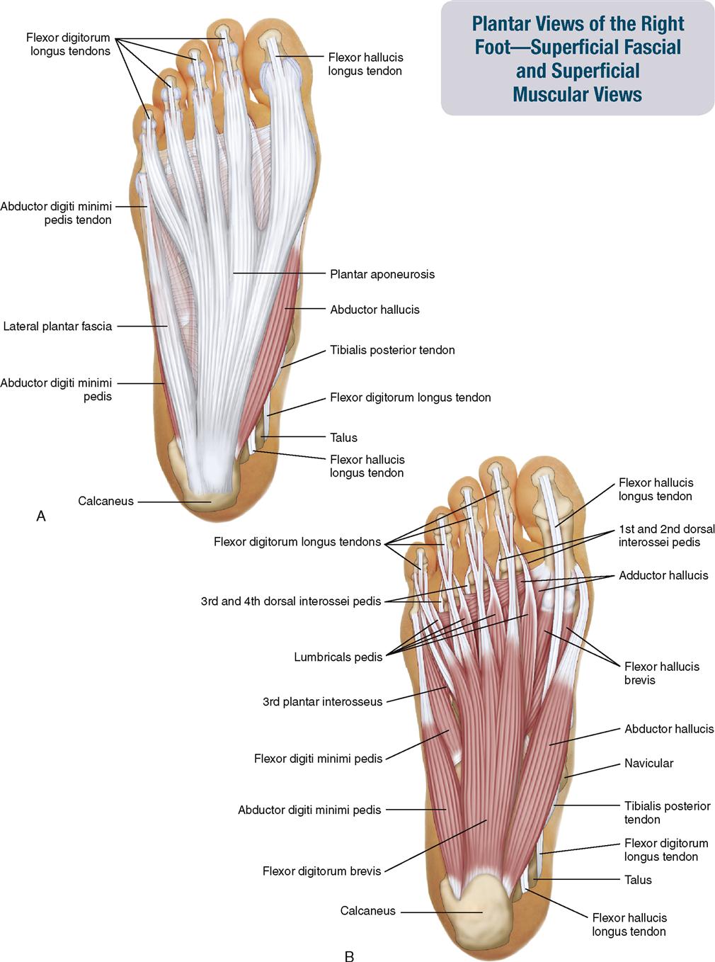
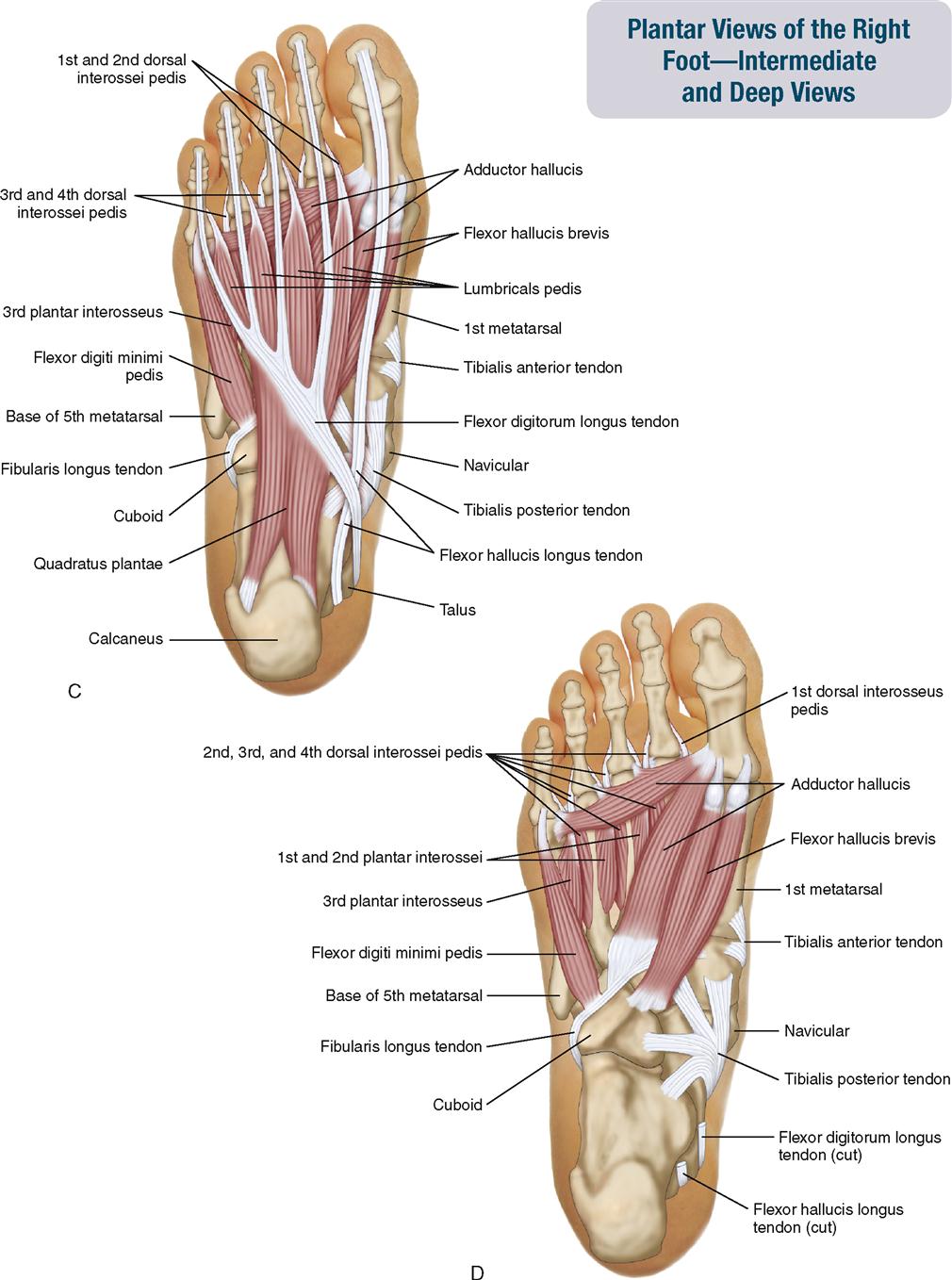
Muscles that move the toes are usually divided into long extrinsic foot muscles and short intrinsic foot muscles. Extrinsic foot muscles originate (attach proximally) in the leg or thigh and insert (attach distally) in the foot. Intrinsic foot muscles originate and insert (attach proximally and distally) in the foot; in other words, they are wholly located in the foot. The intrinsics of the foot are divided into dorsal and plantar muscles. Generally, dorsal muscles extend the toes; plantar muscles flex the toes. The plantar muscles are further divided into four layers, named Layers I through IV, from superficial to deep. The term digitorum refers to toes two through five; the term hallucis refers to the big toe.
The companion CD at the back of this book allows you to examine the muscles of this body region, layer by layer, and individual muscle palpation technique videos are available in the Chapter 11 folder on the Evolve website.
OVERVIEW OF FUNCTION: MUSCLES OF THE ANKLE AND SUBTALAR JOINTS
The following general rules regarding actions can be stated for the functional groups of muscles of the ankle and subtalar joints:


OVERVIEW OF FUNCTION: MUSCLES OF THE TOES
The following general rules regarding actions can be stated for the functional groups of toe muscles:


MUSCLES OF THE LEG AND FOOT
Tibialis Anterior
Pronunciation tib-ee-A-lis an-TEE-ri-or
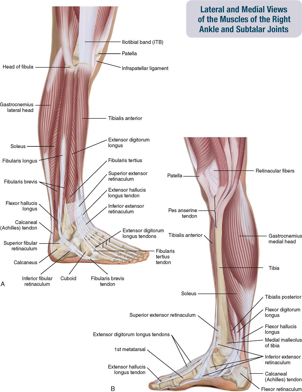
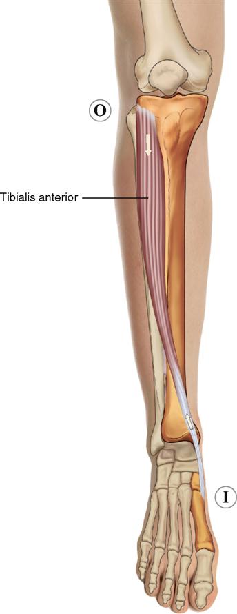
The tibialis anterior is a superficial muscle in the anterior compartment of the leg. It lies immediately lateral to the shaft of the tibia (Figure 11-7).
ATTACHMENTS
Origin (Proximal Attachment)

Insertion (Distal Attachment)

ACTIONS


STABILIZATION
Stabilizes the ankle and subtalar joints.
INNERVATION

PALPATION
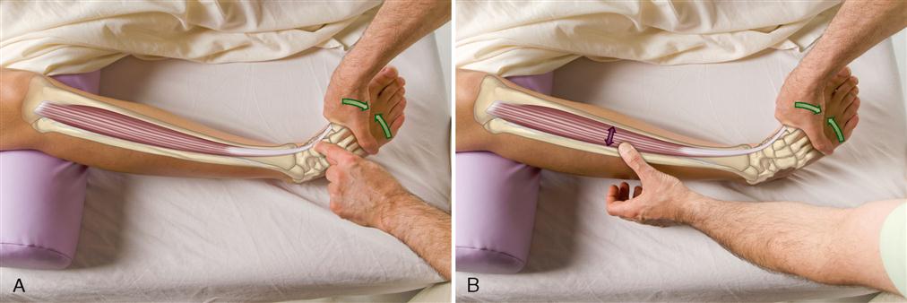
1. The client is supine. Place your resistance hand on the medial side of the distal foot.
2. Resist the client from dorsiflexing and inverting the foot. Look for the distal tendon of the tibialis anterior on the medial side of the ankle joint and foot; it is usually visible (Figure 11-8, A).
3. Palpate the distal tendon by strumming perpendicular across it. Continue palpating the tibialis anterior proximally to the lateral tibial condyle by strumming perpendicular to the fibers (Figure 11-8, B).
TREATMENT CONSIDERATIONS
 The tibialis anterior has a very prominent distal tendon.
 The tibialis anterior and the fibularis longus are known as the stirrup muscles. These two muscles both attach at the same location on the medial foot and may be viewed as a stirrup to support the arch structure of the foot.
 When the tibialis anterior is tight and painful, especially along its tibial attachment, this condition is usually called shin splints or anterior shin splints.
Extensor Hallucis Longus
Pronunciation eks-TEN-sor hal-OO-sis LONG-us
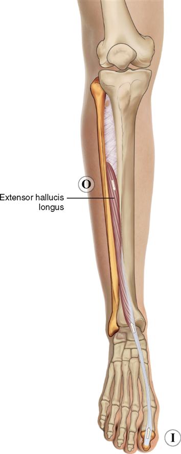
The extensor hallucis longus is a long extensor of the big toe that is located in the anterior compartment of the leg. Most of its belly is deep to the tibialis anterior and extensor digitorum longus, but its distal tendon is superficial as it crosses the ankle joint and on the dorsal surface of the foot (Figure 11-9).
ATTACHMENTS
Origin (Proximal Attachment)

Insertion (Distal Attachment)

ACTIONS
The extensor hallucis longus moves the foot at the ankle and subtalar joints and the big toe at the metatarsophalangeal (MTP) and interphalangeal (IP) joints.


STABILIZATION
Stabilizes the ankle and subtalar joints and the MTP and IP joints of the big toe.
INNERVATION

PALPATION
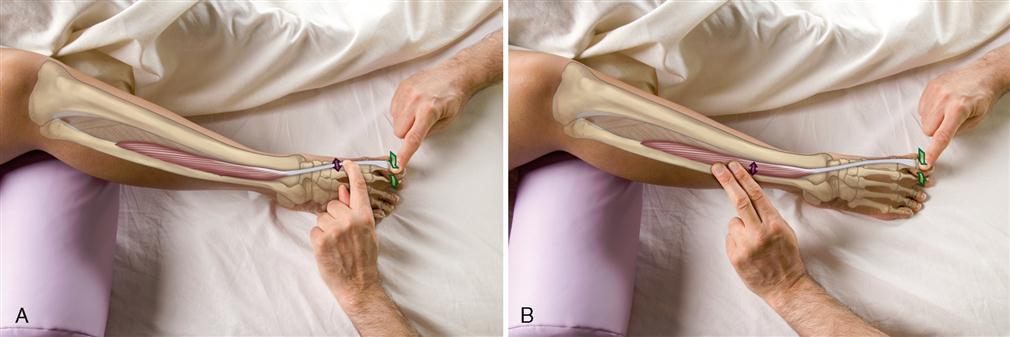
3. Palpate the distal tendon by strumming perpendicular across it (Figure 11-10, A).
4. Continue palpating the extensor hallucis longus proximally. Once it goes deep to the adjacent musculature, do not strum perpendicular to it. Instead, gently place your finger pads over it, and feel for its contraction when the big toe extends (Figure 11-10, B).
TREATMENT CONSIDERATION
When we swing forward during the gait cycle, we usually extend our toes so they do not drag on the ground.

Extensor Digitorum Longus
Pronunciation eks-TEN-sor dij-i-TOE-rum LONG-us
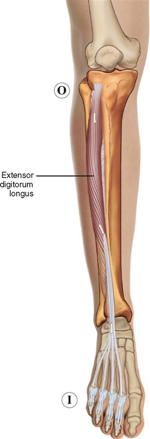
The extensor digitorum longus is a long extensor of toes two through five that is located in the anterior compartment of the leg. Most of it is superficial except for a small aspect of its proximal belly, which is deep to the tibialis anterior and fibularis longus (Figure 11-11).
ATTACHMENTS
Origin (Proximal Attachment)

Insertion (Distal Attachment)

ACTIONS
The extensor digitorum longus moves the foot at the ankle and subtalar joints and toes two through five at the metatarsophalangeal (MTP) and interphalangeal (IP–proximal interphalangeal [PIP] and distal interphalangeal [DIP] joints).


STABILIZATION
Stabilizes the ankle and subtalar joints, and the MTP and IP joints of toes two through five.
INNERVATION

PALPATION
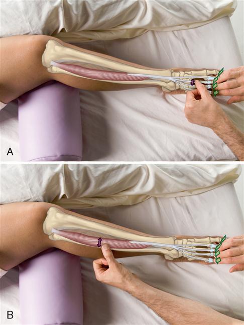
3. Palpate the distal tendons by strumming perpendicularly across them (Figure 11-12, A).
4. Continue palpating the extensor digitorum longus proximally by strumming perpendicular to the fibers (Figure 11-12, B).
TREATMENT CONSIDERATIONS
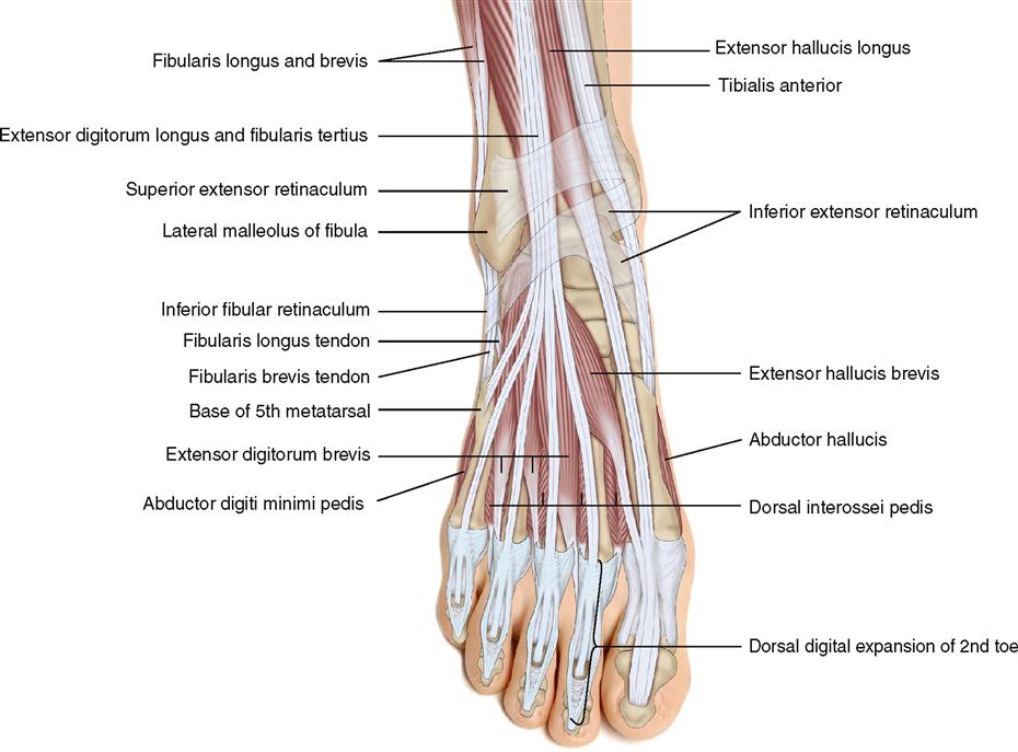
 The distal attachment of the extensor digitorum longus spreads out to become a fibrous aponeurotic expansion that covers much of the dorsal, medial, and lateral sides of the toes. This structure is called the dorsal digital expansion (see page 391) and is an attachment site for many intrinsic foot muscles.
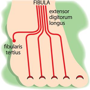
 The most distal and lateral parts of the extensor digitorum longus (which arises from the distal one-third of the fibula) does not attach onto the digits (toes); therefore it is given a separate name, the fibularis tertius.
MUSCLES OF THE LEG AND FOOT: Fibularis Group
Fibularis Longus; Fibularis Brevis; Fibularis Tertius
Pronunciation fib-you-LA-ris LONG-us • fib-you-LA-ris BRE-vis • fib-you-LA-ris TER-she-us
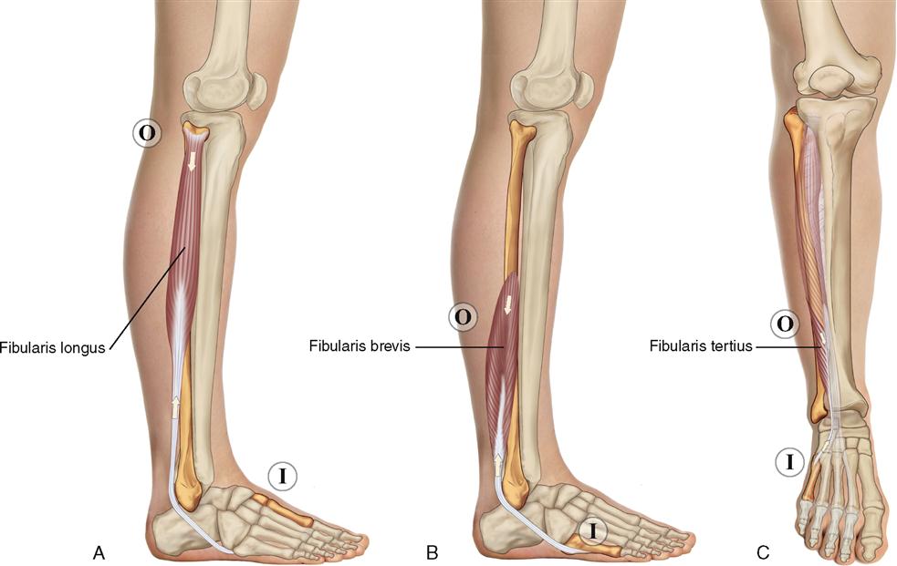
The fibularis group is located laterally on the leg, attached to the fibula. It is composed of the fibularis longus, brevis, and tertius (Figure 11-13). All three fibularis muscles evert the foot at the subtalar joint. The fibularis longus and brevis are located in the lateral compartment; the longus is superficial to the brevis; the fibularis tertius is superficial and located in the anterior compartment. The fibularis muscles used to be called the peroneus muscles.
ATTACHMENTS
Fibularis Longus
Origin (Proximal Attachment)

Insertion (Distal Attachment)

Fibularis Brevis
Origin (Proximal Attachment)

Insertion (Distal Attachment)

Fibularis Tertius
Origin (Proximal Attachment)

Insertion (Distal Attachment)

ACTIONS
Fibularis Longus and Fibularis Brevis


Fibularis Tertius


STABILIZATION
Stabilize the ankle and subtalar joints.
INNERVATION


PALPATION
Fibularis Longus and Fibularis Brevis
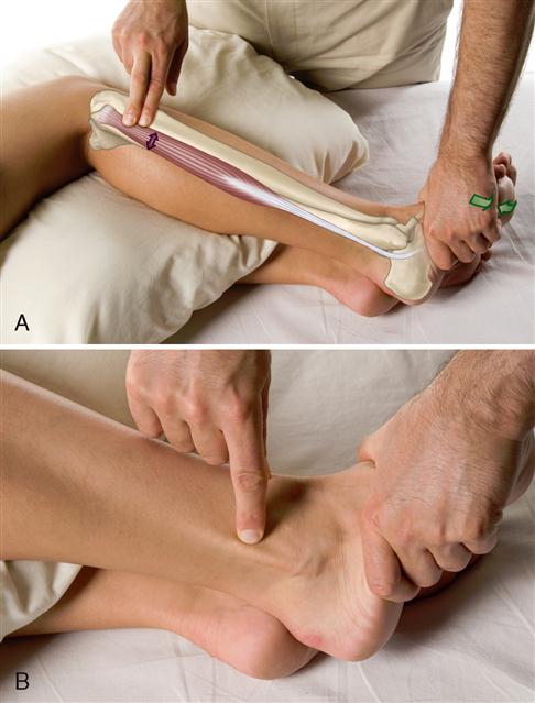
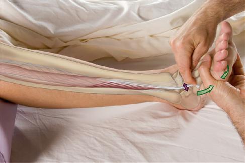
2. Resist the client from everting the foot at the subtalar joint. Feel for the contraction of the fibularis longus (Figure 11-14, A).
3. Continue palpating the fibularis longus distally by strumming perpendicular to the fibers. The fibularis longus becomes tendon approximately halfway down the leg. The distal tendon can usually be seen immediately posterior to the lateral malleolus of the fibula (Figure 11-14, B).
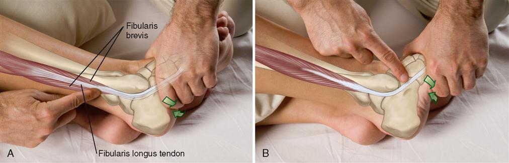
4. To palpate the fibularis brevis, palpate on either side of the fibularis longus in the distal half of the leg (Figure 11-15, A).
5. The distal tendon of the fibularis brevis is often visible and palpable in the proximal foot distal to the lateral malleolus of the fibula (Figure 11-15, B).
Fibularis Tertius
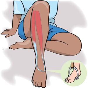
2. If the fibularis tertius is not readily palpable, then resist the client from everting and dorsiflexing the foot and palpate again for its tendon (Figure 11-16).
TREATMENT CONSIDERATIONS

 The fibularis longus and the tibialis anterior are known as the stirrup muscles. These two muscles both attach at the same location on the medial foot and may be viewed to act as a stirrup to support the arch structure of the foot.

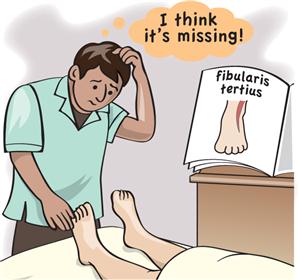
 The fibularis tertius is actually the most distal and lateral part of the extensor digitorum longus. Its fibers do not attach onto a digit (a phalanx); for this reason the fibularis tertius is given a separate name and considered to be a separate muscle from the extensor digitorum longus.

Notes
MUSCLES OF THE LEG AND FOOT: Triceps Surae Group
Gastrocnemius; Soleus
Pronunciation GAS-trok-NEE-me-us • SO-lee-us
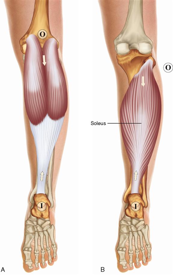
The triceps surae group of the superficial posterior compartment of the leg is composed of the soleus and the two heads (medial and lateral) of the gastrocnemius (Figure 11-17). The gastrocnemius and soleus are grouped together as the triceps surae because they attach together onto the calcaneus via the calcaneal (Achilles) tendon. From the posterior perspective, the gastrocnemius is superficial to the soleus. However, the soleus is superficial in the lateral and medial leg.