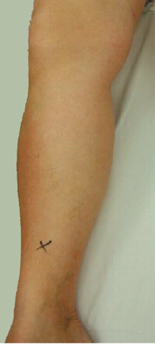
13 Key Points 1. Venous thromboembolism has a serious impact on patients with spinal cord injury. 2. Diagnosis and treatment of thromboembolism are analyzed. 3. Various forms of venous thromboprophylaxis are discussed extensively. Venous thromboembolism (VTE), consisting of both deep venous thrombosis (DVT) and pulmonary embolism (PE), is a leading cause of mortality and morbidity following acute spinal cord injury (SCI) and offer considerable discomfort and delay in rehabilitation for the patients who suffer from it.1,2 SCI has a major global health impact, with a reported annual incidence of 15 to 40 cases per million, and the majority of these injuries occur in young adults from 16 to 30 years of age.3 The frequency of DVT and PE in untreated spinal trauma (spinal fracture with or without SCI) patients has been reported to be between 67% and 100%.4–8 Pulmonary embolism is the third most common cause of death in those patients who survive longer than 24 hours after injury.9,10 The American College of Chest Physicians and the Consortium of Spinal Cord Medicine have recommended specific thromboprophylaxis guidelines for SCI patients.1,11–14 However, routine utilization of thromboprophylaxis measures has great variability due to case-specific risk factors,14,15 low compliance in hospitals,16 questionable efficacy,17 potential complications, and a dearth of high-quality (level I) evidence-based recommendations. In the Spinal Cord Injury Risk Assessment for Thromboembolism (SPIRATE) study, there was a recommendation for more vigorous prophylaxis in SCI patients who are older, obese, and have flaccid paralysis or cancer.18 Moreover, there is uncertainty regarding the length of thromboembolic prophylaxis in the setting of SCI due to questions regarding the role of screening tests for DVT.19 The level of thromboembolic risk is determined by numerous variables. Trauma greatly increases the risk of DVT and PE by causing immobility as well as a hypercoagulable state.11 The risk for VTE appears to be the greatest during the first 12 weeks after injury, when flaccidity, paralysis, and immobilization of the extremities predominate. The pathophysiology of VTE in SCI involves stasis, hypercoagulability, and vessel intimal injury (the Virchow triad)20,21 on the basis of abnormal platelet function and altered fibrinolytic activity in patients with SCI.22,23 Technical factors may also affect the risk of thromboembolic events following surgery.24–26 The method of surgical approach can increase the risk of DVT and PE. Incidence rates are highest for surgeries at lumbar levels and surgeries that require an anterior approach. Moreover, the combination of these two factors increases the risk even further, most likely because an anterior approach to the lumbar spine often requires retraction of the common iliac veins and the venae cavae.24–26 An anterior approach also has an increased risk of pelvic clot formation.24 Other surgical factors that can increase the risk of DVT and PE include length of the procedure, prolonged postoperative immobilization, and prone positioning.25–27 Patient demographics can also contribute to thromboembolic risk. Increased age, male gender, smoking habits, obesity, lower limb fractures, and disease states, such as hypertension, heart failure, diabetes, and cancer, all increase the risk of DVT and PE in patients.1,25–27 Even though spasticity has been suggested to reduce the incidence of thromboembolism, this has not been proven adequately.28 Finally, there exists an association between the occurrence of DVT and heterotopic ossification following traumatic SCI.29 Without prophylaxis, clinical studies using venography as a diagnostic tool for DVT have shown high rates of calf DVT (40 to 80%) in patients suffering from major trauma and SCI.5,27,30,31 Specifically for spinal injuries without prophylaxis, the incidence of DVT reached up to 62%, with SCI and surgery as independent risk factors.5 Proximal embolization has been detected in 50% of established DVT without treatment.32 Death attributed to PE has been found to reach up to 35% of patients with SCI without prophylaxis for thromboembolism.33 The majority of VTE incidents occur in the acute rehabilitation phase of SCI, whereas in chronic SCI, the incidence of DVT/PE is 1.1%/0.3%, respectively.1,34 On the contrary, the incidence of DVT is at least 9.4% in patients with acute SCI who undergo thromboprophylaxis with low-molecular-weight heparin during the acute stage,19 with a mortality rate of 9.7% among SCI patients with DVT/PE.19 Postthrombotic syndrome can lead to prolongation of rehabilitation in 12% of SCI patients with DVT who were followed for 3 years.35 Recurrent leg edema, skin breakdown, pain, relapsing DVT and pulmonary complications appear generally in conjunction with postthrombotic syndrome.33 The gold standard for DVT diagnosis is venography.19 However, due to the invasive character and side effects of this procedure, duplex ultrasonography has replaced venography in diagnosis of DVT.19 Of course, clinical suspicion, together with history/clinical examination (edema of the leg, increase of calf diameter, and localized tenderness along the deep venous system) (Fig. 13.1) and risk factor assessment, are critical to reach the correct diagnosis.36 In mild clinical suspicion of DVT or in cases of technically inadequate ultrasonography, Ddimer measurements within normal limits can exclude DVT.36 Computed tomography (CT) or magnetic resonance imaging (MRI) venography could overcome the technical limitations of ultrasonographic diagnosis of DVT, but further clinical studies are needed to ensure its accuracy.37 However, routine screening for DVT in adults with acute traumatic SCI under thromboprophylaxis is not recommended.19 Fig. 13.1 Clinical photograph of calf edema suspicious for deep venous thrombosis. Similar to DVT, diagnosis of PE can be made by combining history, clinical findings, and diagnostic tests (electrocardiogram, blood gas values, troponin levels, D-dimer values, chest radiography).38 However, objective diagnostic tests for diagnosis of PE include CT pulmonary angiography (CTPA), ventilation perfusion (V/Q) scan, or true pulmonary angiography, with CTPA being the superior of all three.38 Hypercoagulability has been shown to start within a few hours after SCI and persists for at least 2 to 3 weeks.8 Prophylactic pharmacological treatment for thrombosis should, therefore, be initiated as soon as possible after injury,39 and ideally within 72 hours.1 In cases involving intracranial bleeding, hemothorax, intraabdominal bleeding, or other active hemorrhage, pharmacological prophylaxis is contraindicated until the patient is hemodynamically and neurologically stabilized.11 Mechanical forms of prophylaxis, however, may be initiated as soon as possible.1,11 The risk for VTE appears to be the greatest during the first 12 weeks after injury, when flaccidity, paralysis, and immobilization of the extremities predominate.40 The higher incidence of DVT episodes is within the first 2 weeks post-SCI and decreases thereafter.30 Most thromboembolic events occur within 2 to 3 months of injury.41–43 In a retrospective cohort study of 16,240 SCI patients, almost all (88%) thromboembolic events occurred in the first 3 months following SCI.44 Only a few studies have shown occurrence of VTE within the rehabilitation phase or later than 3 months postinjury.45–47 Recent studies report the utilization of thromboprophylaxis for up to 3 months.39,48–50 In a recent meta-analysis,51 the recommended duration of thromboprophylaxis was at least 3 months from the time of SCI or for as long the inpatient rehabilitation period lasts.1 Very few studies suggest the use of thromboprophylaxis beyond 3 months, irrespective of muscle tone state,52 and many support discontinuing pharmacological prophylaxis when lower extremity mobility is purposeful.1,6 In patients with a history of previous DVT or PE, thromboprophylaxis with oral anticoagulants should be extended from 6 months to 1 year depending on the presence of other risk factors.43 Methods of thromboprophylaxis include mechanical methods, pharmaceutical agents, or a combination of the above. Electrical stimulation as a means of VTE prophylaxis has not proven to be efficient.30 Although mechanical prophylaxis with intermittent pneumatic compression devices is a commonly employed method of VTE prevention in SCI patients, its effectiveness has also been questioned.53,54 Pharmacological options include unfractionated heparin (UFH) and low-molecular-weight heparin (LMWH). Mechanical and pharmacological modalities may offer more effective prophylaxis in combination than when used alone; however, this has not been well-established, and a combined approach also raises compliance and cost-related issues.30,55–64 Several studies support the beneficial effect of mechanical prophylaxis alone even in the presence of risk factors (Table 13.1).54,65 The results of a meta-analysis showed no statistically significant reduction in incidence of VTE with the addition of pharmacological prophylaxis.51 Mechanical methods alone provided a quantifiable reduction in VTE,65 and their use is recommended early after injury for at least the first 2 weeks in all patients.1,11 The value of additional pharmacological prophylaxis should be weighed in terms of risks versus benefits specific to each patient. Table 13.1 Risk Factors for Venous Thromboembolism in Spinal Cord Injury Patients Patient Age (> 70 years) Gender (male) Medical comorbidities (cancer, diabetes, obesity, cardiopulmonary diseases) History of deep vein thrombosis and/or pulmonary embolism Smoking status Injury Completeness of SCI (motor complete vs motor incomplete) Other traumatic injuries (head, thoracoabdominal, extremity fracture) Severity of trauma Operative procedure Operative approach (anterior vs posterior) Spinal canal decompression (decompression vs fusion) Number of spinal levels fused (three or more) Level of injury (cervical vs thoracolumbar) A meta-analysis of studies with SCI patients showed that the use of LMWHs was associated with statistically significant fewer episodes of DVT compared with UFH.51 Furthermore, no statistical difference was detected between LWMH and UFH for the prevention of PE.51 This is consistent with a similar meta-analysis in general orthopedic surgery patients.66 Comparisons of LMWH formulations revealed no difference in efficacy between enoxaparin and dalteparin, whereas fondaparinux was more efficacious than enoxaparin.49,65 The use of inferior vena cava (IVC) filters has expanded in trauma patients, including those with SCI. However, objective criteria for their use in SCI patients have yet to be clearly defined. A review of the literature by Johns et al.67 found that IVC filter placement in SCI patients is effective in preventing pulmonary emboli and has a low complication rate. Indications for filter placement include patients who have had pulmonary emboli despite adequate anticoagulation, patients with documented pulmonary embolus who have a contraindication to anticoagulation, patients with concomitant long bone fractures, or patients with free-floating iliofemoral thrombus.67,68 Additionally, filters may be useful in patients with high cervical injuries, poor cardiopulmonary reserve, or IVC thrombus formation despite adequate anticoagulation.67 Only 20% of survey respondents stated that they recommend IVC filters for their SCI patients despite this supporting evidence.69 Bleeding has been found to range between 0.9 to 11% in two previous meta-analyses comparing LMWH and standard heparin in general and orthopedic surgery.66,70 However, the definition of bleeding was heterogeneous across the studies. There is conflicting evidence on the safety of LMWH and standard heparin in studies with SCI patients. In a recent meta-analysis, bleeding was found to be significantly more likely when UFH was used; however, other heparin-related complications were minimal and not statistically different between LMWH and UFH.51 Even though thromboprophylaxis with adjusted-dose UFH produces a cost savings over LMWH,71 the increased bleeding complications associated with UFH and the need for regular monitoring of activated partial thromboplastin time (aPTT) values have led to the use of LMWH in almost all SCI patients according to guidelines of chest physicians, of the American Association of Neurological Surgeons/Congress of Neurological Surgeons (AANS/CNS), and of the Consortium for Spinal Cord Medicine.1,11,13 In addition, there is only moderate compliance with the published evidence-based guidelines on thromboprophylaxis among health care professionals and patients, which needs to be improved.16,72 The cost of thromboembolism in patients with SCI has a major impact on the already high annual cost to the national economy (annual cost to the US economy for SCI patients is $7.2 billion, added cost for thromboembolism $178 million, 1995 data).73 This translates to 1 to 2 weeks’ extension of acute care hospitalization and an increase of costs by 35%.74 Once venous thromboembolism (DVT or PE) diagnosis is established, as soon as possible, intravenous unfractionated or subcutaneous low-molecular heparin treatment should be initiated unless there is a serious bleeding risk. This should be replaced solely by oral warfarin when the international normalized ratio (INR) is within the therapeutic range (2 to 3) and should be continued for 6 weeks to 6 months, depending on the potential benefits and risks for each patient.19 The latest guidelines of the American College of Chest Physicians and the Consortium for Spinal Cord Medicine for thromboprophylaxis in acute spinal trauma, together with recent meta-analyses on the same subject,1,11–13,51 offer the following evidence-based conclusions/recommendations (Table 13.2): 1. Mechanical prophylaxis in both legs should be used for at least the first 2 weeks following acute SCI (level I evidence). 2. Adjuvant pharmacological thromboprophylaxis agents should be initiated within 72 hours of injury if no active bleeding or coagulopathy exists. 3. LMWH is more effective than UFH in DVT prevention in patients with acute SCI; however, PE prevention is equivalent (level I evidence). 4. Fewer bleeding complications were noted when comparing LMWH to UFH in patients with acute SCI (level I evidence). 6. IVC filters are only rarely used in SCI patients who have contraindications to receiving anticoagulation or when anticoagulation is not effective. Table 13.2 Guidelines for Thromboprophylaxis in Spinal Cord Injury Patients Duplex ultrasonography of the deep vein system and a clinical examination are critical for correct diagnosis of deep vein thrombosis Mechanical prophylaxis (at least anti-embolism compression stockings) in both legs should be used for at least the first 2 weeks following acute spinal cord injury Adjuvant pharmacological thromboprophylaxis agents should be initiated within 72 hours of injury if no active bleeding or coagulopathy exists Low-molecular-weight heparin is more effective and safer than unfractionated heparin in deep vein thrombosis prevention in patients with acute spinal cord injury Duration of thromboprophylaxis in patients with complete motor paralysis or in patients with incomplete motor paralysis but with additional VTE risk factors should last for at least 3 months Pearls
Venous Thromboembolism Prophylaxis
 Risk Factors for Venous Thromboembolism in Spinal Cord Injury Patients
 Natural History of DVT/PE in the Absence/Presence of Thromboprophylaxis
 Diagnosis of Venous Thromboembolism
 Start of Thromboprophylaxis in Spinal Cord Injury Patients
 Duration of Thromboprophylaxis in Spinal Cord Injury Patients
 Modes of Thromboprophylaxis in Spinal Cord Injury Patients and Comparisons among Different Modes
Comparison of Mechanical Alone versus Combined Mechanical and Chemical Thromboprophylaxis Methods in Spinal Cord Injury Patients
Comparison of UFH versus LMWH in Acute Spinal Cord Injury Patients
Inferior Vena Cava Filter Insertion in Spine Trauma
 Complications of Pharmacological Prophylaxis in Spinal Cord Injury Patients
 Compliance and Cost-Related Issues of Thromboprophylaxis
 Treatment of Venous Thromboembolism
 Conclusion
 D-dimer measurement within normal limits can exclude DVT.
 Mechanical prophylaxis for DVT of the lower extremities is the essential first measure in all patients with acute SCI.
 LMWH is more effective as DVT prophylaxis than UFH for SCI patients.
Related posts:
 Pharmacotherapy in Acute Spinal Cord Injury: Focus on Steroids
Spontaneous Recovery Patterns and Prognoses after Spinal Cord Injury
Glial Scar and Monocyte-Derived Macrophages Are Needed for Spinal Cord Repair: Timing, Location, and Level as Critical Factors
The Human Central Pattern Generator and Its Role in Spinal Cord Injury Recovery
Spinal and Supraspinal Plasticity after Spinal Cord Injury
Research in Spinal Cord Injury: Building an Effective Translational Research Program
Pharmacotherapy in Acute Spinal Cord Injury: Focus on Steroids
Spontaneous Recovery Patterns and Prognoses after Spinal Cord Injury
Glial Scar and Monocyte-Derived Macrophages Are Needed for Spinal Cord Repair: Timing, Location, and Level as Critical Factors
The Human Central Pattern Generator and Its Role in Spinal Cord Injury Recovery
Spinal and Supraspinal Plasticity after Spinal Cord Injury
Research in Spinal Cord Injury: Building an Effective Translational Research Program
![]()
Stay updated, free articles. Join our Telegram channel

Full access? Get Clinical Tree


Musculoskeletal Key
Fastest Musculoskeletal Insight Engine
