Fig. 8.1
US image of subcutaneous edema on axial plane (asterisk, edema)
Cellulitis
It is a kind of infection that diffuses on the skin and in subcutaneous tissues. There might be injury on the skin. It may be characterized with local sensitivity, pain, edema, and erythema. The most common pathogens are gram (+) aerobic cocci (S. aureus, S. pneumoniae, S. pyogenes) and aerobic and anaerobic gram (−) bacteria. US guides in visualizing the progression of local infection and identifying the necessary interventional procedures [13]. In the US, edematous area (echogenicity increase in heterogeneity) and fibrous connective tissue (increase in hyperechogenicity) become more evident. Increased vascularization pattern helps the diagnosis in CDUS [14].
Cellulitis is treated with antibiotics. In the event that abscess develops, surgical intervention is required. In some cases, an erythematous line, in other words lymphangitis, might extend from cellulitis plaque to the proximal.
Abscess
It is the collection of pus in the dermis or in deeper layers. Majority of the skin abscesses are polymicrobial, and S. aureus is the only factor in 25 % of the cases. Surgical drainage is the most effective treatment in the abscess. Systemic antibiotic treatment is rarely required. US appearance is quite changing (Fig. 8.2). It changes from anechoic fluid collection to irregular hypoechogenicity, to internal echoes, to hyperechoic sediments, to septa, and even to gas formation. Occasionally, only posterior acoustic enhancement is detected. In CDUS, increased vascularization in the abscess wall and adjacent tissues supports the diagnosis. In a PD study, peripheral vascularity increase was observed in 90 % of the cases [15].
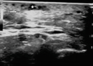
Fig. 8.2
Folliculitis in rectus muscle and edema around it (asterisk, folliculitis; arrow, edema)
Necrotizing Fasciitis
It is a rarely seen progressive soft tissue infection that emerges following minor injuries [16]. Most of the infections are polymicrobial [17]. S. pyogenes is the most common pathogen. Chronic diseases (diabetes and immunosuppressive), IV medication administration, and malignity are among the various factors that increase the predisposition to this disease. The diagnostic value of the US is low at the early period, and fascia gets thicker and fluid collects in deep fascia. The fact fluid collected is measured 4 mm at the thickest part is significant in necrotizing fasciitis [18]. In the advanced cases, the possible abscess and gas formation can be indicated [19].
Early diagnosis, wide-spectrum antibiotic treatment, debridement which cleans all necrotic tissues, regulating liquid-electrolyte balance, regulating the oxygenation of the infected area, nutritional support, and analgesia promotion constitute the basis of the treatment [20].
Trauma
The subcutaneous soft tissue changes that emerge following the trauma might range from minor hemorrhage to fat necrosis, hematoma, and abscess. While the hemorrhage is visualized as a hyperechogenic structure at the early period, fat necrosis is visualized as the hypoechoic areas developed secondary to the necrosis within hyperechoic focus. However, in hematoma, US findings change in time [21]. While it appears to be pseudo-solid at the beginning, it becomes anechoic in time. Within almost a month, hematoma is absorbed. If open wound and abscess formation are observed in hematoma emerged following a trauma, the presence of foreign objects should be investigated. Morel-Lavallee syndrome is the intense seroma that collects between deep subcutaneous soft tissue and fascia in traumas that occur in the lateral side of the upper calf. US illustrates a plenty amount of liquid collection on the echogenic fascia [22]. It is possible to detect the fat necrosis that occurs in this disease and the other affected deep-located structures.
Foreign Body
Foreign bodies might be seen in subcutaneous soft tissue generally following open wounds or perforating traumas. The decision of removing the foreign body is made in line with the size, location, structure, accessibility, and mechanic and inflammatory effects of the object. In case that the pieces that cannot be removed keep staying within the wound and the body cannot figure this foreign object out, a fibrous capsule surrounds it. Inflammation, infection (wound infection, cellulitis, toxicity, tenosynovitis, bursitis, osteomyelitis), and mechanical damage might occur. Foreign object lesions do not have a real neoplasm but are traumatic reactive masses [23]. The sensitivity of the US in foreign bodies is 43–98 % and specificity ranges between 59 and 98 % [24]. It can show wood, bone, sea urchin, and other plantlike objects. A hypoechoic halo like a band occurs around the foreign object in US. It helps it to be differentiated from the neighboring soft tissues. This hypoechoic halo shows increased vascularization pattern in CDUS [25]. Calcification deteriorates the appearance with sesamoid bone and tendon acoustic shadowing. It might be difficult to place probe in places such as interdigital sites, where foreign bodies are found very commonly.
Muscular Diseases
The muscles, which are located around the skeleton, which enable movement, and that we move voluntarily, are skeletal muscles. They make up averagely 40 % of body weight. Skeletal muscle is made up of the combination of hundreds of muscle cells called skeletal muscle fibers with the height ranging between 1 mm and 30 cm. Skeletal muscles have two components, which are muscle fibrils and stromal connective tissue. Fibrils make up fascicles, and fascicles make up muscles. Since the skeletal muscles are structures that can extend from one joint to another, long linear probes are used when they are examined. Two adjacent muscle tissues appear as hyperechoic band formation (Fig. 8.3).
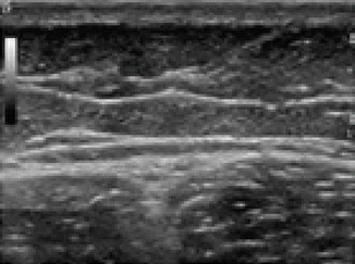
Fig. 8.3
US image of normal muscle tissue on axial plane
While muscles are visualized as homogenous, multiple, thin parallel echoes on the longitudinal plane, they are more irregular, dispersed thin echoes on the transverse plane. Bright echogenic connective tissue fascia is present around the muscle.
The pathologies of muscles can be assessed with US. These are strain/tear, hematoma, myositis ossificans, myositis, compartment syndrome, rhabdomyolysis, hernia, and tumors [26].
US can detect the presence, the location, and the severity of the anatomic damage. US demonstrates the volume increase, edema, or fluid-pus collection in a detailed manner [27] (Fig. 8.4).
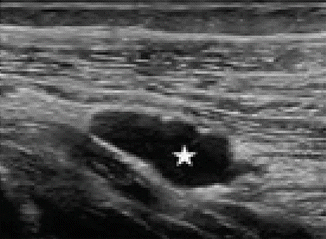
Fig. 8.4
Cyst within the rectus muscle (star)
Trauma
Trauma constitutes the biggest part of the muscle pathologies. Different situations might emerge according to the cause and location of the lesion. In a minor tear, mild heterogeneity or local hematoma might occur in the muscle tissue, and in the big lesions in myotendinous junction, hematoma might extend to the external fascia (Fig. 8.5).
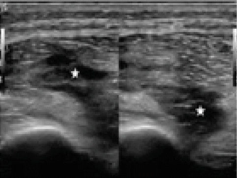
Fig. 8.5
Dispersed hematoma areas secondary to muscle tear (star)
Muscle damages might have extrinsic or intrinsic causes. Myotendinous junction is the location where partial or complete muscle tears are most commonly seen (Fig. 8.6). Tears might also occur in compartments close to the myotendinous junction.
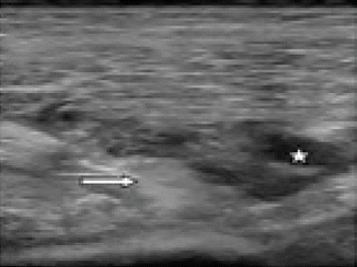
Fig. 8.6
Edema and hematoma areas secondary to muscle tear (star, hematoma; arrow, edema)
Type II muscle fibrils located at both sides of the joint and helped with the high energy and rapid movements in the main muscles have higher probability of tearing [28]. Rectus femoris, biceps femoris, and medial head of gastrocnemius muscle are the most commonly affected muscles (Fig. 8.7).
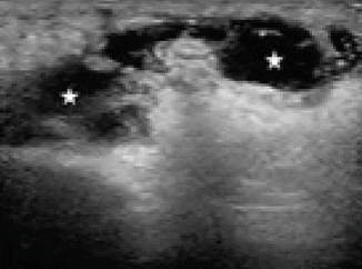
Fig. 8.7
Large hematoma in rectus femoris muscle secondary to rupture (star)
US can assess the separation and retraction of muscle fibrils from tendon or aponeurosis. Four to 6 weeks are needed for complete healing following the trauma. The fibrous tissue that emerges in the late period appears as hyperechoic areas within the muscle in the US [29]. Residual fibrous tissue scar and recurrent injury risk are directly proportional. One of the rarely seen complications in muscle tear healing is cyst formation within the muscle. Septa formations might be visualized.
Muscle dissection is visible on fascial planes between the muscles. In more severe injuries, intramuscular fluid collection is visible. Big hematomas can cause compartment syndrome with mass effect [30].
Muscle hematoma might be seen in different ways in the US according to the stage. It might contain septa. Intramuscular hematomas are not different from the hematomas in any part of the body. While the active hemorrhage appears hyperechoic, it might be hypoechoic within a couple of hours. Later on, liquid self-leveling might be seen. After a few days, the collection becomes anechoic in a uniform way. These collections are absorbed within several weeks.
Contusion is visualized in fibroadipose septa following direct trauma. This septum is seen as prominently thick in US. Diffuse hemorrhage results in increased echogenicity. Intramuscular hematoma results from the damage to the epimysium vessels.
Muscle contraction and fibrous septa-fascicle relationship can be presented with dynamic US. In case of contraction, while muscle tissue is visualized as thick and hypoechoic, interfibrillar septa can be visualized in different ways.
In children, contusion coexistent with intramuscular bleeding and crush in muscle tissue without damaging the skin integrity directly secondary to the trauma following sports injuries is the most common. Damage occurs to the fibrils and intramuscular hematoma accompanies. In US, pathology is commonly visible in the location where trauma occurs. Thus, it is distinctive from myotendinous tears by appearance.
Primary Pyomyositis
It is a subacute deep bacterial infection of the skeletal muscle mainly seen in tropical regions [31]. Infection is widely visible in quadriceps, gluteal, and iliopsoas muscle and the most common causing factor is S. aureus [32, 33].
In addition to the chronic conditions such as immunodeficiency or diabetes mellitus, factors such as malnutrition contribute to its development. Also, secondary pyomyositis might arise in this case [32]. On US, diffuse hyperechogenicity is seen in the involved muscle at the inflammatory phase [13].
While pyomyositis responds to antibiotic treatment at the early phase, drainage is the best treatment option when abscess develops. In CDUS, the finding showing the progress of lesion into abscess is the formation of limited mass with a thick wall at various echogenicity between hypoechoic and hyperechoic, where increased vascularization is visible in the wall or internal septa [15].
Myositis Ossificans
It is a benign lesion described with nonneoplastic heterotopic bone formation in the soft tissue. It results from intramuscular hematoma calcification or ossification. Patients apply to the hospital with a painful hard mass that occurs within the soft tissue and continues to grow. At the beginning, within the first 3 weeks after the trauma, a disorganized, nonhomogenous soft tissue mass is seen on US. At this phase, it is not possible to differentiate the soft tissue tumor with US. Three to 4 weeks after the trauma, calcification becomes visible. These calcifications mainly have peripheral distribution and eventually, ossification occurs. In the course of progression, acoustic shadowing helps the radiologist in making the diagnosis. The absence of abnormal soft tissue distribution enables the distinction from parosteal sarcoma. Normal periosteum adjacent to cortex excludes parosteal sarcoma.
US has an important role in the diagnosis of muscle tumors. The nature (cystic/solid), location (local/diffuse), vascularization pattern, size of the lesion, and its relationship with the adjacent soft tissues can be visualized. In malign tumors, irregular boundary, heterogeneous echo, structural deformities in the adjacent structures secondary to the infiltration are seen. Benign tumors have regular boundary, homogenous echo structure. Also, displacement occurs in adjacent tissue.
Tendon Diseases
Tendons connect the muscles to the bones. They help the bones move by transferring the load of the muscle to the bone. Water constitutes 70 % of the wet weight of the tendon. The most visible cells in the tendon are fibroblasts. Tendon is made up of Type I collagen fibrils. Fibrils combine and make up fibers and fibers combine and make up fascicles, which contain blood, vessels, and nerves. This structure helps the tendon to stress and stretch [34].
US is the primary option in tendon pathologies because it allows for dynamic examination. It should be performed along with both longitudinal and axial planes. In longitudinal examination, tendons appear as hyperechoic bands similar to stria, with uniform thickness, and as thin parallel fibril echoes. In transverse examination, round or oval echoic spots with homogenous distribution are visible.
Synovium is visualized as thin hypoechoic frame at the anterior and posterior sides of the tendon. Paratenon (fibrous connective tissue) is visualized as ambiguous hyperechoic tissue that continues with subcutaneous fatty tissue.
Tendon pathologies include tendinosis and partial tear, tendon dislocation, full-thickness tears (Figs. 8.8 and 8.9), inflammatory processes (tenosynovitis and peritendinitis), tendon tumors, and postoperative findings.
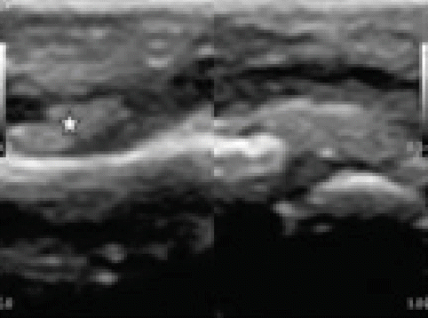
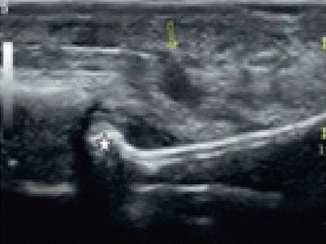
Fig. 8.8
Total tear in the tendon (asterisk)
Fig. 8.9
Flexion contracture secondary to the total tendon tear in hand (arrow, total tear; star, contracture)
Tendonitis
Tendon swelling, irregular fibril appearance, local intratendinous hypoechoic areas, and calcifications are seen. In abnormally thick tendons, increased blood flow pattern, and neovascularization are detected in CDUS. It is assumed that these changes show aging and tear probability. This is because degeneration, micro tears, and interstitial tears in the tendon can provide similar US findings that complement each other and that can be visualized concurrently [30].
Longitudinal hypoechoic areas are visualized in intratendinous small tears. In case that there is no coexisting hematoma, it is difficult to differentiate from tendonitis. It might be accompanied by degeneration and small fractures. It might be finalized with the healed small tear degeneration. While no volume loss is visible in tendinitis, defect with good boundaries is seen even in the partial tears.
In full-thickness tears, diagnosis is easier with US compared to partial tears. This is because the thickness of the tendon fibers is degenerated totally, the torn edges of the tendon are separated, granulation tissue and hypoechoic hematoma are visible, tendinitis is seen in tendon ends, irregular contours emerge in tendon, and posterior shadowing is visible in the torn edges.
The diagnosis might get difficult in case that the edema that develops in the acute period or tendon that tears in some cases is compensated with another tendon. The absence of tendon retraction and broad defect in the partial tear is an important finding in differentiating it from the full-thickness tear. Dynamic examination is required for making the final diagnosis in tendon tears. It is possible to stress the tendon with mildly passive moves and make it visible [35].
In full-thickness tears accompanied by degenerative diseases in adults and in full-thickness tears secondary to the trauma in children, rupture is observed at the location where tendon attaches the bone [36].
Achilles is the thickest and the strongest tendon in human body. It is made up of the combination of tendinous parts of gastrocnemius and soleus muscles. Achilles tendon rupture is one of the most commonly seen tendon tears. Mostly, it occurs (44–83 %) during exercise and is more widely seen among men than women. Since vascularization is weak, the rupture generally occurs in this 5–7 cm distal of the tendon. On US, normal tendon appearance disappears due to hematoma formation. It is possible to evaluate the tendon thickness with US.
Inflammatory processes of the tendon can be evaluated between a wide range of simple tendinosis and advanced-stage tendon pathologies. On US, thickening in tendon, heterogeneity, and focal hypoechoic areas are seen. On CDUS, increased flow signal is visible in and around the tendon.
An overwhelming majority of the tendons, excluding some tendons like Achilles and plantaris, are covered with a synovial sheath. Therefore, pathologies of the tendon and synovial pathologies are observed.
Tendinitis
Tendinitis refers to tendon inflammation. Etiopathogenesis of the tendinitis is very wide. Trauma, partial rupture, and metabolic and rheumatismal disorders lead to inflammation. Besides, inflammation might arise out of mechanic factors following tendon sheath irritation.
< div class='tao-gold-member'>
Only gold members can continue reading. Log In or Register to continue
Related posts:

Stay updated, free articles. Join our Telegram channel

Full access? Get Clinical Tree


