Fig. 8.1
Aphthous ulcer on the lower lip shows a fibrinous base

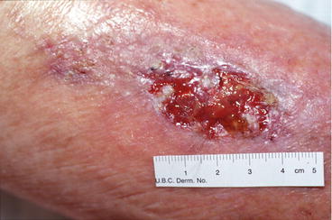
Fig. 8.2
Right leg image shows pyoderma gangrenosum with an undermined ulcer
Reactive arthritis or Reiter’s syndrome (RS) is triad of reactive arthritis and conjunctivitis following urogenital or gastrointestinal tract infection. Patients with RS may have superficial painless oral and genital ulcers. The genital ulcers in the male have a characteristic of circinate white plaques that grow centrifugally on the uncircumcised glans penis (circinate balanitis).
Systemic lupus erythematosus (SLE) . Digital ulceration is a rare manifestation of SLE. It occurs most commonly in patients with disease of long duration and may be the result of vasculitis, the presence of antiphospholipid antibody, or atherosclerosis. In addition to digital ulcers, sometimes panniculitis and vasculitis can lead to ulceration. Lupus panniculitis occurs on the face, the breast, the abdomen, and thighs and is seen in 2–5 % of SLE patients. Vasculitis may cause lower leg ulceration.
Systemic sclerosis (SSc) . The most common type of ulceration in SSc is the digital ischemic lesion (DIL). These ulcerations occur in up to 40 % of patients with SSc and can be extremely debilitating due to pain. The lesions come in two forms: those occurring on the digital pulp and those occurring overlying extensor joints (such as the interphalangeal joints or metacarpophalangeal joints). The digital pulp lesions usually occur on the digital tips, although they can occur at the hyponychium (the junction of the free edge of the nail plate and the fingertip) or on the lateral tips of the digit. They present as punctate ulcerations filled with a hyperkeratotic inclusion without surrounding erythema and are usually quite tender. They resolve with a pitted scar. The other type of lesion overlying the extensor joints typically presents as true ulcerations, which can often be associated with some crust and/or eschar. They often heal with lacy telangiectasias. In addition to being found on the hands, they are also seen overlying the elbows, knees, and malleoli. These lesions are likely the result of a combination of trauma and poor blood supply.
Another cause of ulceration in the SSc patient is pyoderma gangrenosum. These commonly present as ulcers on the leg with a rolled, inflammatory border and they can change in size quickly.
Get Clinical Tree app for offline access

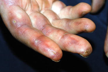
Dermatomyositis (DM). An ulcer in DM can be the result of calcinosis or vasculopathy. In general, ulcers can indicate one of three scenarios: first, necrotic skin ulceration, especially in non-acral regions and regions without surrounding rash, is considered a predictive factor for the presence of underlying cancer; second, ulceration of Gottron’s papules can occur following DMARD therapy, especially methotrexate or mycophenolate mofetil; and third, patients with melanoma differentiation-associated gene 5 antibodies (MDA5) have a higher incidence of ulcerative cutaneous lesions on the digital pulp and periungual areas, within Gottron’s papules, and over the elbows (Fig. 8.3) [1]. In addition diffuse punched-out ulcerations and ischemic digital necrosis have been described in literature. These patients may be at higher risk of interstitial lung disease. It should be noted that ulcers can also occur in areas of intense inflammation and may be of no particular prognostic significance.

Fig. 8.3
Right hand in dermatomyositis patient. Note the ulceration on the digit pulp and the lateral erythema
Stay updated, free articles. Join our Telegram channel

Full access? Get Clinical Tree


