Tumors


Tumors


Malignant tumors of the musculoskeletal system constitute 10% of new cancers in children, numbering approximately 1,000 cases in the United States per annum. Benign tumors are 10 times the rate. We are presently on an exponential curve for advancement in oncology, for example, overall 5-year survival has risen from 10% to 20% in 1970 to better than 70% today. The most common pædiatric tumor is the benign fibrous cortical defect. The most common malignant tumor is osteosarcoma of bone and rhabdomyosarcoma of soft tissue; Ewing sarcoma traverses tissue types. This chapter focuses on common diagnoses.


EVALUATION

Because the word “tumor” may have sinister connotations, consider classifying tumors in simple terms for patients [A]. Most tumors are not life threatening. They may threaten local tissues, for example, risk of morbid fracture, and thus require surgical management. They may be stable, amenable to conservative measures, or incidental findings, which are left alone.


Physical Examination

Take the temperature. Ewing sarcoma may be mistaken for osteomyelitis at first. Observe gait for alteration due to pain or other interference with function. Does the child look ill, as if overcome by pain or a generalized process? Are the complaints focal or diffuse? Are there “hard” objective signs? Are the soft tissues reactive, as evidenced by swelling, redness, induration, and adhesion? Is there articular stiffness to suggest guarding? Is there atrophy of disuse or other asymmetry? If a mass is detectable, is it tender? Is it soft, firm, or hard? How large is it? Greater than 5 cm is ominous for soft tissue sarcoma.


Imaging

Develop a method that is clear, standard, efficient, and reproducible.

Röntgenogrammes The acronym ALLMDS facilitates communication [D]:


  • A: Age.


  • L: “Looks like.” This admits descriptives such as “sunburst,” “moth eaten,” scalloped,” and “expansile” [E].


  • L: Location. Where within the bone, for example, metaphysis, diaphysis, or epiphysis [F], or what bone, for example, long bone such as the femur, irregular bone such as the spine, or flat bone such as the pelvis [G].


  • M: Margins. These are divided into distinct, called “geographic,” or nondistinct, called “nongeographic.” The latter is more ominous, as it is a sign of rapid tumor growth and failure of bone to react and delimit this growth.


  • D: Density. Lesions may be lytic, which are radiolucent; blastic, which are radiodense; or mixed, which are heterogeneous. This aids local assessment of bone but is not prognostic. Lytic lesions erode the integrity of bone and risk morbid fracture, influencing decision on internal fixation. On the other hand, while blastic lesions may be more stable, they may represent a high-grade osteosarcoma.


  • S: Soft tissue. This may be reactive, such as elevation of periosteum, or a mass, for example, Ewing sarcoma.


D Imaging for bone tumor Simple algorithm.


E Some diagnostic features by röntgenogramme.


F Location of tumors within a long bone.


G Tumor types by skeletal site Common location of benign tumors in green. Malignant tumors in red, and common sites indicated in bold.

Ultrasonography This is indicated for a soft tissue mass when there is a low index of suspicion for malignancy. It may be used as a guide to aspiration or needle biopsy. It is simple to perform in an awake child without a special facility or medication.

Scintigraphy This is a measure of bone turnover and activity of a lesion. It may discern between a benign lesion that is incidental and not the explanation for regional pain and one that has a microfracture, thereby guiding treatment. It surveys the skeleton for other sites of disease. It has a high negative predictive value for malignant disease. Overlap between benign and malignant disease reduces the positive predictive value.

CT This is essential to staging as a survey of the chest and abdomen. Its fine bone detail aids operative decision making.

MRI Like CT, MRI is essential to staging. It is indicated for soft tissue tumors and bone tumors with a significant soft tissue component. It is supplanting CT for bone, for example, osteoid osteoma of a long bone. It has high sensitivity but specificity declines in differentiation of tumor from infection. The principal disadvantage is the requirement for sedation in the young child.

PET Detects gamma rays from the positron-emitting radioactive isotope fluorine-18 substituted for a hydroxyl group on glucose (fluorodeoxyglucose). The glucose analogue is taken up by actively dividing cells, where it is trapped but not metabolized, resulting in concentration of radioactive signal in tissue with high glucose uptake. This modality may enhance CT and surveys for metastases and is useful to follow response to treatment.


Laboratory Analysis

A peripheral blood smear is useful in leukæmia. White blood cell count may be difficult to interpret, for example, it may be reduced due to chronic disease. Inflammatory markers may be increased in tumor, for example, Ewing sarcoma, or in infection. Elevation of alkaline phosphatase may be sensitive though nonspecific, for example, in the active phase of fibrous dysplasia. It may serve as a marker of response to medical treatment, such as with bisphosphonates. Blood cultures distinguish infection. Special tests may be indicated, such as urinary catecholamines and metabolites, which are elevated in neuroblastoma.


Staging

There are three stages of benign tumor.


  • Latent. These may be asymptomatic and incidentally found. They show little or no growth and do not disturb or escape the compartment.


  • Active. There is growth of tumor, as well as destruction, remodeling, and possible fracture of bone, limited to the confines of the compartment.


  • Aggressive. Tumor grows rapidly; destroys, distorts, and fractures bone; and escapes the compartment into surrounding soft tissue, including rare metastasis (e.g., giant cell tumor).


Staging of malignant tumors is complex and disease specific. It is essential to management and prognosis of malignant tumors. Several factors contribute to stage [H]. Surgical staging (Enneking) emphasizes pathologic grade and the compartmental nature of a lesion: fascia, joint, and bone define a compartment [I]. Broadly, stage 0 may be considered precancerous. For stage I, surgical treatment is sufficient. Surgery for stage IV is prophylactic or palliative, for example, for impending or actual morbid fracture. Stages II and III combine medical and surgical management.


Biopsy

This may be percutaneous or open. Percutaneous may be of bone marrow from the iliac crest, for example, in leukæmia or Ewing sarcoma, or with image guidance when open access is morbid, for example, in the spine, or when there remains uncertainty about the diagnosis of tumor. Plan a large and wide enough specimen from multiple locations to be definitive. Biopsy may be incisional for diagnosis, excisional when geographic and localized, or compartmental when margins are indistinct but there is no regional or distant spread [J]. The principles of open biopsy are well established, founded on the spirit that this must be undertaken with the same level of preparation as the definitive procedure.


  • Plan for future reconstruction when selecting incision, which should be as small as possible and extensile.


  • Perform a transmuscular approach to limit contamination within that compartment.


  • Avoid major neurovascular structures. Unlike muscle, these are not expendable in the event of future resection.


  • Include the margin of a lesion, where growth and atypia tend to be greatest.


  • Strict hæmostasis reduces contamination.


  • Intraoperative frozen section confirms that enough tissue has been obtained to establish diagnosis or that a lesion is benign before proceeding with definitive care.


  • Consider referral before biopsy if the surgeon, pathologist, and institution are not equipped to manage the case regardless of diagnosis.


Differential Diagnosis

The adage “culture tumor, biopsy infection” was conceived in the diagnostic dilemma each can pose the other. Consider fever, systemic signs, age, location, and elevation of inflammatory markers. Presence or absence of a mass and appearance on imaging sharpen the diagnosis [K]. Biopsy may be necessary for certainty.

Distinguish osteosarcoma from myositis ossificans by zone reversal. The latter is characterized by central proliferating cells surrounded by a margin of ossification. In osteosarcoma, the rapidly growing periphery has not yet ossified.

Differentiate fractures by history, for example, trauma or repetitive stress, and empirically by treating with rest and immobilization. Distinguish anxiety time from biologic time: 4 to 6 weeks will not alter tumor prognosis. Laboratory analysis is normal. Advanced imaging may be necessary, such as MRI showing no soft tissue extension or mass. Resist biopsy.


I Staging of musculoskeletal tumors Surgical staging is determined by grade and physical extent of lesion.


Factor


Comment


Tumor


1: confined to site of origin


2: outside the site of origin


Nodal involvement


0: none


1: local


2: regional


3: distant


Metastasis


0: absent


1: present


Grade


1: low


4: high


Serum markers


Elevation


Resection


Measure of free or contaminated margins


Vascular


Invasion of vasculature


Lymphatic


Invasion of lymphatic system


Response


To chemotherapy, based upon % necrosis in surgical specimen


Imaging


e.g., MRI


H Some factors considered for staging of tumors These may vary according to disease type.


J Biopsy of tumor This may be incisional, within the tumor (red), excisional (white dots), or compartmental (green dots).


K Infection, not tumor Atypical features of infection include periosteal reaction and diaphysial location (red) and extension from metaphysis transphysis to epiphysis (yellow).


1


2


3


Pain


Mild


Moderate


Functional


Location


Upper limb


Lower limb


Proximal femur


Size


< 1/3


1/3-2/3


> 2/3


Density


Blastic


Mixed


Lytic


A Assessing risk for morbid fracture of hole in bone 3 points are given for pain that is sufficiently severe to disturb function. A critical value is 9, which represents the beginning of the exponential part of the fracture curve (>1/3 will fracture). This system is a guide: it was developed for bone metastases in adults (Mirels).


B Active unicameral bone cyst There is a fracture fragment floating inside the cyst like a “fallen leaf” (yellow). The fracture takes up the entire width of the bone and has eroded the cortex to the point of fracture (red).


C Aspiration and injection of a bone cyst Injection may be of steroid, bone marrow aspirate, or other adjuvant.


BONE CYST


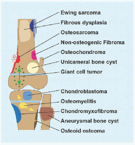
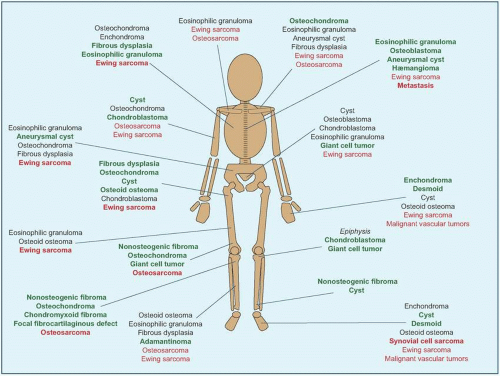
Unicameral Bone Cyst

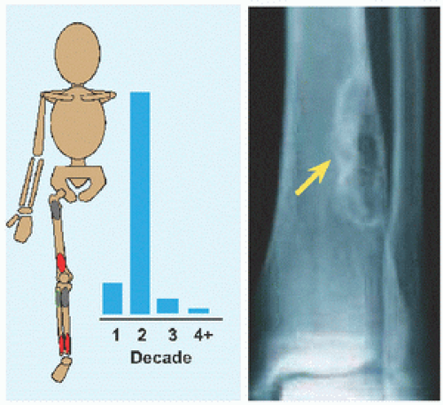
The name implies a “single chamber” (Latin camera = Greek καµαρα: “vaulted chamber”) without loculation, although some have septæ. This most commonly occurs in the humerus and femur. Cause is unknown. The cysts are filled with yellow fluid and lined with a fibrous capsule.

Evaluation These may be incidental or may present with pain. They are metaphysial and travel toward the diaphysis with growth. Incidental findings are assessed for fracture risk [A]. Pain indicates bending under load or fracture and may be severe enough to interfere with function. Lesions in the proximal femur are high risk because of concentration of the force of weight bearing and the grave anatomic consequences of fracture in this region.

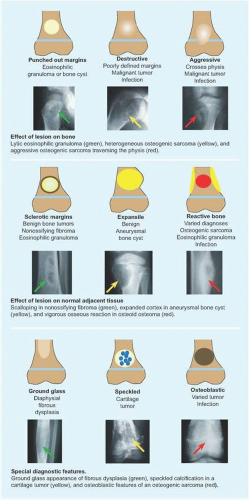
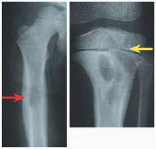
RÖNTGENOGRAMME The lesion is central, nonexpansile, geographic with a sclerotic margin, and lytic. It may have septæ and an osseous fragment may float in its midst, known as “fallen leaf sign.”

Management The natural history is spontaneous resolution of cysts by maturity in most cases. Latent cysts may be observed. Active cysts, which abut the physis, grow inexorably, and may fracture, tend to be treated surgically. Age, location, and fracture risk and consequences guide treatment.

LOW RISK EXCEPT FEMUR Observe. A humeral cyst that is small, asymptomatic, or acceptably painful may be managed conservatively, such as activity modification and nonnarcotic analgesics, and may resolve with maturity.

LOW RISK PROXIMAL FEMUR Observe or treat surgically because of concerns regarding load at, and fracture of, the hip.

HIGH RISK Observe if patient is very young and humerus is involved, or treat surgically. The proximal femur trumps age: consider fixation even in the very young.

FRACTURE EXCEPT FEMUR Treat by closed methods as long as alignment is acceptable. Healing of the fracture may partially heal cyst, potentially converting a cyst that is high risk by size into one that is low risk due to filling in with new bone.

FRACTURE FEMUR Treat surgically. Immobilization, for example, in hip spica cast, is associated with residual deformity, in particular coxa vara, due to intrinsic instability of the fracture and incomplete healing. For the proximal femur, improved alignment with surgical fixation may reduce complications of fracture such as malunion and osteonecrosis.

Surgical treatment may be divided into cases.

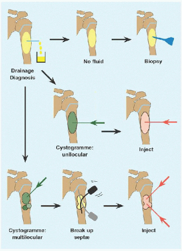
PERCUTANEOUS WITHOUT FIXATION This began as injection of steroid (Scaglietti), for its angiostatic and fibroblastic inhibitory effects. The method has evolved in multiple directions such that there is no consensus on best practice. It is preferred for the upper limb and lower risk lower limb lesions.


  • The cyst is drained. If it is solid, reconsider diagnosis and perform an incisional biopsy.


  • A cystogramme determines whether the cyst is uni- or multilocular. If the former, inject with steroid or other adjuvant. Bone marrow aspirate may bring mesenchymal stem cells that will promote bone ingrowth and healing. Calcitonin inhibits osteoclasts. Calcium sulfate cement is osteoconductive. Proprietary fibrosing agents have been promoted.


  • If the latter, break up septæ to form a unilocular cyst. In the process, perforate the cyst to create channels of communication with the medulla, based upon the rationale that altered hæmodynamics with venous obstruction may be a causative factor, and to allow access to the cyst by bone stem cells.


  • Repeat for recurrence.


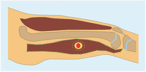
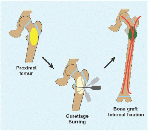
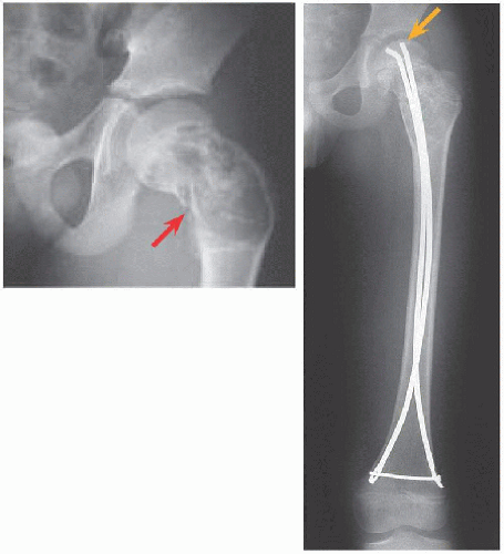
OPEN WITH FIXATION This is indicated for high-risk lesions, such as proximal femur [D], or where open access is simple, for example, calcaneus.


  • Create an ovoid window to avoid stress concentration at an angle. The cyst may dictate location of this at the eroded cortex.


  • Débride the wall to remove all cells, manually or with power.


  • Consider adjuvant intralesional therapy to kill residual cells, for example, liquid nitrogen (freezing), phenol (chemical), argon laser (coagulation), or hydrogen peroxide. For liquid nitrogen, leave in cavity until it evaporates; for other chemicals, let sit for 2 to 3 minutes. Cycle thrice. Reduce application hazard by controlling the agent within the osseous cavity in order to minimize injury to surrounding soft tissue. These augment but do not substitute for complete débridement.


  • Pack the cyst with graft, allogenous or autogenous, or with a substitute. There are no robust data suggesting anything is better than bone, and allogenous bone avoids the morbidity of harvesting bone, in particular because much may be needed to fully fill a large cyst.


  • If the bone is stable, apply a cast. If internal fixation is indicated, for example, proximal femur, medullary implants will load share and thereby be more durable in the event of recurrence. In the immature child, elastic medullary nails may be inserted antegradely or retrogradely. They provide stability even in proximal lesions because they may be anchored in the unaffected and hard proximal epiphysis: no physial arrest will occur given their smooth surface and small fractional cross-sectional area [E].

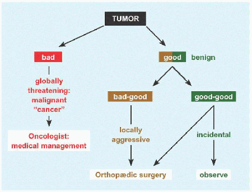
Complications While bone cyst is a benign or “good” tumor, it is a “bad-good” tumor because outcomes can be vexing.


  • Recurrence. Some factors cannot be controlled, such as abutment against a physis. The principal factor that is controllable is débridement; hence the recommendation to use a burr, to be methodical, to proceed until healthy bleeding bone, and to leave only periosteum in places (so long as the bone is not unreasonably destabilized or resected).


  • Growth disturbance. This may be iatrogenic or related to activity of cyst. The former may be minimized by avoiding physis during operation. The latter is a reflection of duration and aggressiveness of cyst.


  • Malunion. This is most common in the proximal femur. Likelihood increases with closed management.


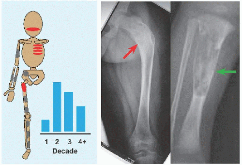
Aneurysmal Bone Cyst

As the name implies, this is vascular and expansile. Cause is unknown, although vascular malformation is most subject to speculation. Evaluation and management principles overlap with unicameral cyst, with key distinctions. Aneurysmal cyst is more aggressive:


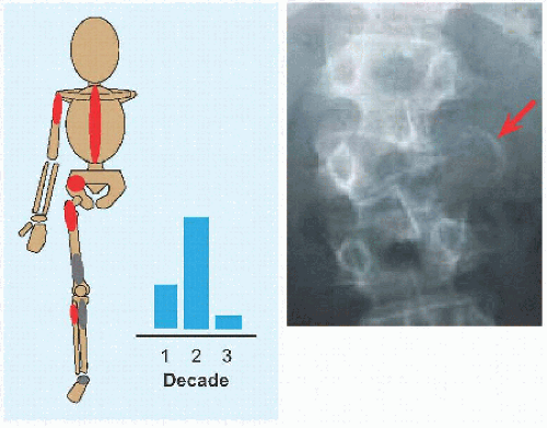
  • Location. 1/4 arise in the axial skeleton, including posterior elements of the spine and the periacetabular region of the pelvis [F]. Consequences are greater morbidity of disease, for example, neural compromise, and of treatment, including more invasive dissection and more complex reconstruction.


  • Recurrence. Correct surgical technique has reduced a formerly universal rate. Recurrence occurs within 2 years of operation, although children must be followed until maturity.


  • Association with other tumors, including giant cell and osteosarcoma, in which it may represent the cystic component of a primary tumor.


D Open treatment of bone cyst The cyst is débrided and bone grafted (green), after which the femur is stabilized with elastic medullary nails (red).


E Open treatment of bone cyst This cyst fractured (red). Retrograde elastic nailing was stable by purchase in the hard, unaffected proximal epiphysis (orange).


F Distribution of aneurysmal cyst The axial skeleton, including the spine (red) and the pelvis (white), makes advanced imaging and open treatment more typical.


G Aneurysmal cyst of the pelvis The expansile and otherwise aggressive appearance on röntgenogramme led to MRI, which shows a characteristic blood-fluid level (yellow).


A Nonosteogenic fibroma It peaks in adolescence and commonly affects the tibia. It is eccentric, loculated with a scalloped margin (yellow).


B Fibrous dysplasia The femur is distorted to resemble a “shepherd’s crook” (red). Note the thick periosteal healing response. Appearance may be so varied, including provoking concern (green), that further imaging often is necessary.

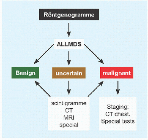
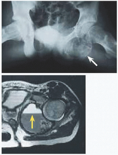
Evaluation On röntgenogramme, it is eccentrically expansile, lytic, and multiloculated, giving a “soap-bubble” appearance. The thin margin has been likened to an “eggshell.” The radiographic appearance may raise concern and prompt further imaging. Scintigramme may show a “halo” of increased uptake surrounding a central cold region. MRI will show blood-fluid levels [G] and define soft tissue extension.

Management This tends to be open and has the additional considerations:


  • Preoperative angiography and selective embolization before surgical treatment of large lesions, in particular in the pelvis, will decrease hæmorrhage and may shrink the lesion to decrease recurrence where complete resection is unrealistic.


  • Entry into a cyst will return blood. Include soft tissue within, or the lining of, the cyst for biopsy, which will show proliferative fibroblasts, spindle cells, osteoid, and multinucleated giant cells.


  • Break up all septæ to define the entire cyst wall, which may be interrupted.


  • Consider an en bloc resection with reconstruction where feasible to reduce recurrence.


  • In the spine, excision may result in instability necessitating reconstruction, including bone grafting, fusion, and internal fixation.


BENIGN FIBROUS TUMORS


Nonosteogenic Fibroma

Originally reported as a cyst of bone (Phemister), this was named separately as nonosteogenic (Jaffe) and nonossifying fibroma. The appellation fibrous cortical defect is reserved for small lesions confined to the cortex without medullary extension.

This is the most common benign lesion of bone in children, peaking in adolescence [A]. Cause in unknown, although it has been related to physis and traction by tendon or ligament, for example, at origin of the gastrocnemius from the distal femur.

Evaluation Most are incidental findings on röntgenogrammes obtained for injury followed by pain.

RÖNTGENOGRAMME The lesion is metaphysial, eccentric, geographic with a “scalloped” margin, and lytic with one or more loculations [A]. A large lesion may show a cortical breach.

SCINTIGRAMME This is indicated when there is uncertainty whether the lesion is a source of pain, suggesting structural failure, or when the lesion risks morbid fracture, such as in the proximal femur or >50% diameter of the bone. Fracture will demonstrate increased signal.

Management The natural history is spontaneous resolution by maturity. Observe incidental findings and follow clinically based upon symptoms. For fracture through a lesion, immobilize and observe for (partial) healing. If the fracture cannot be immobilized effectively, or for lesions with significant risk of morbid fracture, treat like a unicameral cyst (q.v.).


Fibrous Dysplasia

Neoplastic fibrosis replaces and weakens bone, causing microfractures that hurt and lead to progressive deformity. It may arise in any bone, though the ribs and the proximal femur [B], as well as maxilla, are distinctive sites. Two types are distinguished: monostotic (80%) or polyostotic. The latter subtype is more severe and occurs in conjunction with café au lait spots and precocious puberty as part of McCune-Albright syndrome (cf. Syndromes chapter) and in conjunction with soft tissue myxomata in Mazabraud syndrome. Polyostotic fibrous dysplasia is caused by an activating gain of function mutation in the GNAS1 gene located on chromosome 20q13.2. This results in abnormal synthesis of both organic and inorganic components of the extracellular matrix, thereby compromising the structural integrity of bone.

Only gold members can continue reading. Log In or Register to continue

Stay updated, free articles. Join our Telegram channel

Aug 18, 2016 | Posted by in ORTHOPEDIC | Comments Off on Tumors

Full access? Get Clinical Tree

Get Clinical Tree app for offline access