Neuromuscular Disorders


Neuromuscular Disorders


Neuromuscular disorders account for the greatest burden of chronic disability in children. Because motor dysfunction is often an early manifestation, the orthopædic surgeon may be the first to evaluate the child.

Injury to the central nervous system may include several outcomes [A]. The prevalence of neuromuscular disease is evolving [B]. Poliomyelitis has declined due to immunization, and spina bifida has declined due to dietary supplementation with folic acid during pregnancy. Cerebral palsy remains unchanged, in part because advances in obstetrical knowledge and practice are balanced by increased survival of premature infants.

Neuromuscular disorders may be distinguished based upon level of disease [C]. Such a simplified system serves as a basis upon which to organize the approach to complex patients.


EVALUATION


Physical Examination

The neural examination includes:


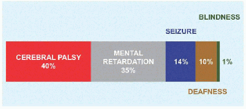
  • Cognition. Cognitive function is essential to evaluation, management, and outcomes. For example, it may be difficult to determine pain in a cognitively impaired child with cerebral palsy.


  • Motor function. Assess strength manually and against body weight. In addition to individual muscle groups, a functional assessment is based upon standing and walking ability: standing for demonstration, standing to assist transfer, walking at home, and walking in the community. For walkers, determine total distance as a general measure of function. Assistive devices are not limiting but liberating.


  • Sensory function. Altered sensation may be direct, due to neural loss, for example, spina bifida, or indirect, due to cognitive loss.


  • Special signs, for example, a child using the hands to “walk up” the anterior legs and thighs in order to rise from a seated position in the setting of muscle weakness. Divide tone into normal, reduced (hypotone), or increased (hypertone).

Gait Evaluate by observation (cf. Lower Limb chapter). Gait is divided into stance and swing phases, which overlap for 20% of the normal cycle. Stability during single-limb stance can be limited by cognition, balance, proprioception, coordination, standing posture, bony deformity, contractures, and weakness. Without stability in stance, it is impossible to develop an effective gait pattern. Each swing phase requires clearance of the off-loaded foot to preposition that foot in terminal swing. Ankle equinus, weakness in dorsiflexors, and cospasticity are examples that will prevent clearance and prepositioning and lead to an ineffective gait, one with inadequate stride length, poor cadence, or otherwise biomechanically impaired and energy inefficient. Gait also may be assessed in a laboratory, including instrumented motion analysis, dynamic electromyography, pedobarography, and energy consumption test. Include walking and running, which as a stress test will amplify deficit, for example, asymmetry of the limbs in hemiplegia.

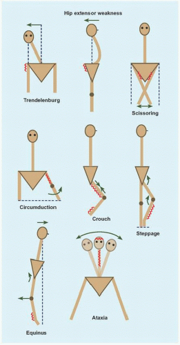
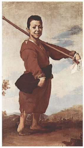
There are several morbid gaits [E].

HIP ABDUCTOR WEAKNESS This produces a Trendelenburg gait, which is characterized by shifting of the center of mass over the affected joint in stance to eliminate the moment arm. Because the patient has the tendency to fall away from the weak limb in stance, a compensatory mechanism is to raise the walking velocity, thereby reducing time for the effect of gravity, for example, advanced hip deformity.

HIP EXTENSION WEAKNESS Walking slows to reduce forward momentum. Lumbar lordosis increases to move center of mass posteriorward. Knee flexion decreases to limit hip flexion, for example, muscular dystrophy.

SCISSORING Adductor spasticity slows walking velocity and narrows the base of stability, for example, diplegic cerebral palsy.

QUADRICEPS WEAKNESS This reduces knee control and is the principal determinant of walking. Body weight flexes the knee, which is counteracted by hip extension, plantar flexion, and locking of the knee in extension. Additionally, the limb may be rotated lateralward to move the force vector medialward away from the sagittal plane, for example, spina bifida.

CIRCUMDUCTION GAIT Hamstring weakness, or quadriceps contracture or spasticity, reduces knee flexion, which is a major hindrance during swing. The limb is functionally lengthened, necessitating that it be swung outward for the foot to clear the ground.

CROUCHED GAIT This may be compensatory to reduced hip extension, due to knee flexion contracture, or a result of triceps suræ weakness. The gait cycle is shortened, there is forward trunk lean to reduce demand on quadriceps, and energy consumption increases, for example, overlengthening of tendo Achillis.

STEPPAGE GAIT Anterior crural muscle weakness is compensated for by increasing hip and knee flexion for swing phase toe clearance.


D Select milestones.


E Abnormal gait Site of disease in red and motion, compensatory and morbid, in green.


F Effect of time on contracture formation The orange part of the curve may be lengthened by early intervention such as stretching and bracing.


G Some contracture tests These are common tests to assess contractures in cerebral palsy.


H Pelvic obliquity Rotate and neutralize the pelvis to unmask a primary spine deformity (red) that persists despite neutral alignment of pelvis and hips or a primary hip deformity (green) that remains despite pelvic and spine position. Combined deformities are present when pelvis rotation has no influence.

EQUINUS GAIT Reduced ankle flexion leads to a toe-toe gait, concentrating force at the forefoot during stance. There is reduced flexion or extreme hyperextension of the knee. In the neuromuscular patient, with compromised proximal muscle strength and control, forward translation of the center of mass reduces the effective base of support, thereby reducing stride length, for example, muscular dystrophy.

HINDFOOT VARUS This concentrates force over the lateral border of the foot during stance, for example, Charcot-Marie-Tooth disease.

ATAXIC GAIT Greek (α-: “not,” τα§ιζ: “precise arrangement”). This is a sign of central nervous disease, for example, in the cerebellum. It is characterized by a broad base, short stride, and titubation due to impaired balance.

The neuromuscular patient must balance benefits against energy consumption of walking. Speed declines first, to maintain energy cost per time. However, energy cost per distance increases: most patients will select a wheelchair for mobility once this exceeds thrice the normal.

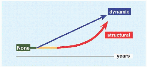
Deformity Deformity may be dynamic or static.

DYNAMIC This reflects errant neural signaling. It may vary by position, for example, upright posture increases tone that accentuates dynamic deformity. Surgical correction is compensatory and less predictable.

STRUCTURAL Dynamic deformity may become fixed or structural with time [F], such as due to spasticity or positioning in wheelchair. Manual interventions before structural deformity sets in, such as stretching and bracing, may delay onset. Sites of contracture include the skin, muscle, and joint capsule or ligaments. Surgical correction is direct, but may be limited by concomitant contracture of neurovascular structures, which would be intolerant of stretch, for example, popliteal artery and tibial nerve behind a flexed knee. With more time, fixed contractures lead to joint deformity and instability, for example, flattening of the head of the femur subluxated against the rim of acetabulum in cerebral palsy.

Deformity may develop even in the setting of hypotonia, for example, scoliosis. Contracture matters when compensatory mechanisms are overwhelmed, thereby interfering with function. Titrate expectations and interventions according to function. For example, a child in a wheelchair has less demand than does an ambulatory child; by contrast, a plantigrade foot is a universal goal.

HIP FLEXION This tips the trunk forward, which is compensated for by lumbar hyperlordosis or knee flexion into a crouched gait. Assess this by the prone extension test [G] or by extending the affected hip in the supine position with the opposite hip maximally flexed to eliminate compensatory lumbar lordosis (Thomas). 30 degrees is a guide to release.

HIP ABDUCTION Hip abduction is important for perineal access and care. While limitation of abduction <45 degrees is a guide to release, let function be the ultimate guide.

ILIOTIBIAL TRACT The patient lies decubitus with affected limb up and with opposite hip and knee flexed. Flex, abduct, and extend the affected hip to bring tract over the trochanter major, where it will be tensioned to prevent the ipsilateral knee from adducting beyond midline under gravity (Ober).

GRACILIS In the prone position, abduct the hip with the knee flexed: tensioning the biarticular gracilis by extending the knee causes hip adduction (Phelps).

RECTUS FEMORIS This biarticular muscle flexes the hip and extends the knee. In the prone position, flexion of the knee tensions the muscle: contracture is revealed by the elevation of the buttock as the hip is obligatorily flexed (Duncan-Ely). Rectus femoris contracture also may limit knee extension and draw the patella proximalward (alta).

KNEE FLEXION Measure this by the popliteal angle. Hip flexion to 90 degrees tensions the hamstrings, thereby exposing contracture as the knee is extended.

KNEE EXTENSION Genu recurvatum is abnormal. It may be a direct result or a compensatory mechanism for weakness at the knee.

EQUINUS Invert the hindfoot to lock the subtalar joint. Flex and extend the ankle in knee flexion (gastrocnemius relaxed) and knee extension (gastrocnemius tensioned). 10 degrees of flexion allows heel-toe gait.


0 degree allows a plantigrade foot in the nonambulatory. >30 degrees risks crouch gait in the setting of weakness.

ROTATIONAL PROFILE This often is abnormal in neuromuscular patients. Medial femoral torsion is a characteristic of cerebral palsy. Lateral tibial torsion is a characteristic of spina bifida.

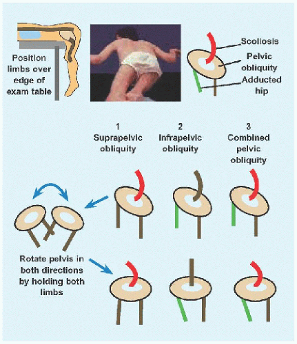
PELVIC OBLIQUITY This may be suprapelvic, originating in the spine, or pelvic, due to hip deformity [H].

SPINE Deformity is common in neuromuscular disease. Assess this in the unweighted prone position, upright, and with traction. Pay attention to the skin for signs of decompensation, such as sore, due to limited movement, and for surgical incisions, for example, spina bifida repair.

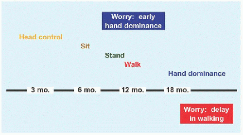
Reflexes Knowledge of reflexes, reactions, and signs is essential to understanding normal and delayed development [I].

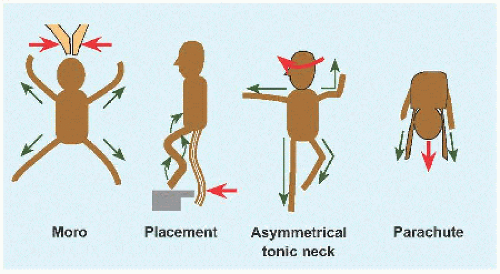
MORO A startled baby, for example, simulated fall or clapping, abducts and extends all limbs and spine, which may be followed by opposite movement into an embrace [J]. The reflex is lost by 6 months. This aids differentiation of neonatal paralysis, for example, brachial plexopathy, from pseudoparalysis, for example, due to clavicle fracture.

PLACING REACTION In the vertical suspension position, the anterior leg is brought into contact with an edge: the normal infant flexes hip, knee, and ankle to surmount the edge and spontaneously extends the lower limb when the sole is planted. The reflex also may be elicited in the upper limb with the dorsal forearm as initial point of contact. This is normal up to 12 months.

ASYMMETRIC TONIC NECK Supine and neutral neck. Turn head in one direction then the other: the limbs toward which the head is turned extend while the opposite flex, assuming a “fencer position.” This is lost by 6 months.

PARACHUTE Suspend the baby prone by holding the waist. Simulated fall elicits extension of the upper limbs toward and to protect the head. This appears at 6 months and remains.

EXTENSOR THRUST In the vertical suspension position, pressing the soles down against a flat surface elicits hip and knee extension for support. This reflex is lost by 6 months. Persistence will interfere with normal reciprocal hip and knee flexion during swing phase.

VERTICAL SUSPENSION A baby suspended vertically by the axillæ with examiner’s thumbs supporting the neck flexes the hips and knees until 6 months. Extension and scissoring are signs of spasticity.

PALMAR GRASP The digits contract to receive an object inserted into the palm, and tone in the entire limb increases as the object is withdrawn. This reflex is lost by 6 months.

BABINSKI Plantar stimulation results in extension of the hallux and fanning out of the lesser toes. This reflex is lost by 2 years.

TONIC LABYRINTHINE In the supine position, tilting the head backward produces opisthotonos. This reflex is lost by 6 months.

DEEP TENDON Corticospinal reflex to acute muscle stretch. Hyperreflexia, including clonus >5 beats, indicates upper motor neuron disease.

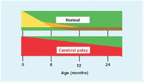
While primitive reflexes aid diagnosis of developmental delay, they also are prognostic: loss of these reflexes by 2 years is predictive of independent walking.

Imaging

MRI This is the modality of choice for evaluation of the brain. It shows congenital malformations, such as polymicrogyria, heterotopia, and schizencephaly. Other findings in developmental delay include intracranial hæmorrhage or ischæmia, periventricular leukomalacia, cystic encephalomalacia and ventriculomegaly. It also allows determination of whether myelination is appropriate for age.


I Natural history of reactions, patterns, and reflexes. Most reflexes are lost by 6 months (yellow) and others by 12 months (orange). Persistent primitive reflexes and pathologic patterns represent a delay in development, such as in cerebral palsy (red). Appearance of the parachute reflex (brown) at 6 months and its persistence are normal.


J Common reflexes. Provocation in red and response in green.


Study


Value


Total lymphocyte count


> 1500/mm3


Albumen


> 3.5 g/dL


Transferrin


> 175 mg/dL


K Laboratory analysis before operation. These values are associated with reduced infection rate.


Laboratory analysis The diagnosis of neuromuscular disease is typically not dependent upon laboratory analysis.

CYTOGENETICS Chromosomal analysis for a genetic disorder.

INFECTION Because of the high prevalence in this patient population, tests such as urinalysis often are indicated. Infectious workup also screens the mother for in utero exposure.

NUTRITION Preoperative assessment may reduce infection rate after major operation, for example, spine fusion for cerebral palsy [K].

PULMONARY FUNCTION Preoperative testing estimates risk of prolonged ventilator dependence after spine fusion in muscular dystrophy.

ELECTROENCEPHALOGRAPHY Diagnosis and evaluation of seizure.

ELECTROMYOGRAPHY AND NERVE CONDUCTION Diagnosis of muscular dystrophy and neuropathy.


A Cognitive dissonance In Æsop’s fable, the fox decides that the grapes he cannot reach are not ripe yet but sour, thereby reconciling his desire with his inability to fulfill it. In surgery, this may set into motion a cycle in which family perceives benefit despite equivocal outcomes, which confirms both their and the surgeon’s choice of treatment, perpetuating the process.


A Causes of cerebral palsy.


Tone


Comment


Pyramidal


Spasticity


Velocity-dependent hypertonicity 80%


Extra-pyramidal


Athetosis


Greek αθετοζ: “unable to position” Slow, writhing, convoluted


Choreiform


Greek χορεια: “dance” Sequential, rhythmic, ballistic


Ataxia


Imbalance, incoordination, tremor


Mixed


Heterogeneity of disease


B Tonal classification of cerebral palsy Spasticity is caused by loss of inhibition of the reflex arc. Velocity dependence is manifested by tone that is dependent on the rate of stretch. Spastic patients have hyperreflexia, clonus, and myostatic contracture. Many patients will defy distinct classification. Response to surgical intervention is most predictable in spasticity.


Site


Features


Hemiplegia


Affects half the body in sagittal plane (Greek ‘εµι: “half”) Focal intracranial hæmorrhage or infarct


Diplegia


Affects “both” lower limbs (Latin bis: “both”) Periventricular leukomalacia due to vascular insufficiency


3/4 are premature


Tetraplegia


Affects “four” limbs (Greek τετταρεζ: “four”) Variable cognitive dysfunction, including total involvement characterized by diffuse brain disease.


Monoplegia


Rare


Mixed


For example, asymmetric diplegia, double hemiplegia


C Geographic classification of cerebral palsy.


MANAGEMENT PRINCIPLES

Balance the burden of disease with the morbidity of intervention. Orthopædic care addresses the downstream effects of a primary disease that it cannot cure. Prioritize function over deformity: “normal” is unrealistic and unattainable. Prioritize communication, independence, and mobility over walking.

Appreciate the significance of sensation and perceptive disabilities. Terms such as “spasticity” and “plegia” do not acknowledge sensory impairment in cerebral palsy.

Account for all costs: pecuniary, social, and familial. Think of the family as a computer: running too many and incompatible programs may crash it.

Beware of cognitive dissonance. This describes the tension that results from attempting to reconcile two contradictory beliefs or realities, a tension that may be relieved by changing one of the two to be consistent with the other. To parents and caregivers with high expectations before operation, a poor or even fair outcome may be dissonant whereas a good outcome will be harmonious. This may explain how a positive subjective outcome is not contradicted by a negative objective outcome. An understanding of cognitive dissonance may bridge the divide between family expectations and surgeon restraint.

Recognize that natural history may be harnessed when favorable, such as in nonprogressive conditions or those with spontaneous improvement, yet it can be an obdurate force against influence, hence the failure to preserve walking despite the best intentions and surgical interventions.

Adhere to the proof razor: the burden of proof lies with one who makes a claim and not with one challenged by it. Exhaustive and exhausting remedies, foisted upon vulnerable families, may be rejected absent evidence. Do not be dogmatic: absence of proof is not proof of absence. Alternative remedies are acceptable when they do not harm the child or family; when they do not delay, interrupt, or otherwise interfere with proven treatments; and when there are no other effective options. Recognize the ethical limitations of caregivers consenting to high-risk procedures on behalf of a cognitively impaired patient. Be mindful of the ethical imperative primum non nocere: “first do no harm.”


CEREBRAL PALSY

The name describes motor dysfunction caused by a disease of the brain. The suffix -plegia is derived from Greek πληγη: “a blow, stroke,” used by Hippocrates to describe “paralysis” resulting from being struck by disease. While it is referred to as static encephalopathy, in recognition of a defined insult resulting in a stable brain disorder, the musculoskeletal consequences are progressive. It also is known as Little disease, after the English physician W. J. Little (1810-1894), who championed tenotomy (having undergone the procedure himself) and who attributed the disease to difficult pregnancy, premature birth, and neonatal asphyxia.

The appellation encompasses a group of disorders resulting from pre- and perinatal brain insult as well as postnatal causes such as near drowning and traumatic brain injury [A]. 1/4 walk independently, 1/2 have capacity with walking aids, and 1/4 do not walk.


Classification

The disorder is classified according to tone [B] and geographically [C]. Pyramidal pathways (in particular corticospinal) directly continue to the motor neurons of the spinal cord. The extrapyramidal system, including the basal ganglia and cerebellum, indirectly modulates motor function. A lesion in this system results in “dyskinetic” movements, characterized by abnormalities in control, posture, and timing. The geographic classification correlates with timing and mechanism of brain injury [D]. For example, the basis for prematurity as a risk factor is incomplete development of the cerebrum. Vasculature, resulting in hypoperfusion to the periventricular white matter, the region most susceptible to injury
because it is a watershed zone between striate and thalamic arterial systems. These areas carry fibers responsible for lower limb motor function, manifesting as spastic diplegia. Geographic subtypes have several characteristics [E]. A final component of classification is the Gross Motor Function Classification System [F].


Medical Considerations

Multiple comorbidities beyond the musculoskeletal system impact decision making and outcomes.

Cognition Half of patients with cerebral palsy have cognitive impairment. This correlates with severity. It is exacerbated by dysarthria, which impedes expression of intellectual capacity.

Skin Hygiene is difficult, impeded by contracture, and dependent on caregivers. Skeletal distortion without the ability to accommodate due to movement restriction may lead to decompensation such as decubitus ulcer.

Gastrointestinal Poor oromotor control has several consequences:


  • Failure to thrive due to feeding and swallowing difficulty. Patients may require gastrostomy or jejunostomy to augment nutrition. Low weight is the greater concern, by contrast with spina bifida, where overweight challenges function.


  • Gastroœsophageal reflux and associated aspiration pneumonia.


  • Constipation.

Teeth Dental caries, enamel dysgenesis, and malocclusion.

Respiratory Aspiration pneumonia due to oromotor dysfunction and seizure. Motor dysfunction impairs cough. Reduced mobility reduces mechanical factors that aid lung inflation. Prolonged recumbency after operation adds risk.

Seizure 1/3 of patients have epilepsy, a direct result of brain insult. This correlates with extent of involvement, increasing with cognitive impairment and tetraplegia.

Visual Visual field defects due to cortical injury. Premature infants may develop retinopathy.

Hearing Loss is seen in patients with history of kernicterus.


Movement Management

This may be focal, to address local or segmental spasticity, or it may be generalized.

Botulinum toxin This neurotoxin, produced by the Gram-positive anaerobic rod Clostridium botulinum (Latin botulus: “sausage,” after poisoning from contamination), binds to motor nerve terminals where it cleaves SNARE proteins to inhibit release of acetylcholine. It first was used to treat strabismus. It is injected into the skeletal muscle to produce a dose-dependent reversible paresis to overcome spasticity and potentiate manual lengthening. Needle placement may be determined anatomically or aided by ultrasonogramme or electromyography.

Botulinum toxin may be administered therapeutically or diagnostically. It is most effective in the lower limb, in particular triceps suræ in combination with casting for equinus. Other applications include the hip adductors for scissoring, to reduce subluxation of femoral head, and to facilitate perineal care, the rectus femoris to alleviate stiff knee gait, and the upper limb for flexion deformities of elbow and wrist. It may serve as
an adjunct to preoperative assessment of tendon lengthening, providing a reversible preview of the operative effect. Because it blocks dynamic muscle activation, it is not effective for fixed contracture.


D Timing of brain insult Although correlations are imprecise, the earlier the event during gestation, the greater the injury.


Features


Hemiplegia


Diplegia


Tetraplegia


Disability


Mild


Moderate


Severe


Feet


Equinovarus, equinovalgus


Knees


Mild


Moderate


Severe


Hips


OK


Subluxation


Dislocation


Spine


OK


OK


Scoliosis


Upper Limbs


Variable


Little


Major


Seizure


Common


Rare


Common


Walking


Yes


Variable


No


E Characteristics of geographic subtypes Boy with a clubfoot by Jusepe de Ribera (1591-1652) is a vivid depiction of hemiplegia. The boy stands in equinus, with the forearm pronated and the wrist and fingers flexed.


Level


Function


I


Walking: without limitation.


Running and jumping.


Decreased speed, balance, coordination


II


Walking: with limitation For example, railing for stairs.


Difficulty on uneven surface or interactive environment


III


Walking: at home with hand-held mobility device Community: wheel-chair.


IV


Does not walk.


Stands to transfer.


Supported sitting.


Wheel-chair for mobility.


V


Global impairment.


Unable to independently walk, stand or sit: requires transportation


F Gross Motor Function Classification System Because it measures selfinitiated movement and ability, this is a functional assessment, as opposed to systems that describe type of movement or part of body affected. It consists of 66 measures of sitting (truncal control) and walking ability, subdivided according to age.


Maximum dose of botulinum toxin is 20 U/kg, distributed as 2 U/kg in small muscles and 6 U/kg in large muscles, and 600 U total. Maximal effect is seen in 1 to 2 weeks. Duration of effect is 3 to 6 months; redosing should be no more frequent, in order to avoid immunity.

Phenol This also is known as carbolic acid, after its discovery from coal tar. It was the agent used by the Scottish surgeon Sir Joseph Lister (1827-1912) to clean wounds and soak bandages in his development of antisepsis. It targets a major motor nerve directly (rather than diffusing over muscle endings), which is identified with a nerve stimulator under general anæsthesia. It is most effective for musculocutaneous nerve to improve extension of the elbow and obturator nerve to improve hip abduction.

Only gold members can continue reading. Log In or Register to continue

Stay updated, free articles. Join our Telegram channel

Aug 18, 2016 | Posted by in ORTHOPEDIC | Comments Off on Neuromuscular Disorders

Full access? Get Clinical Tree

Get Clinical Tree app for offline access