and Hans-Christoph Pape2
(1)
Department of Traumatology, University Hospital RWTH Aachen, Pauwelsstraße 30, Aachen, 52074, Germany`
(2)
Department of Traumatology, University Hospital RWTH Aachen, Pauwelsstrasse 30, Aachen, 52074, Germany
Abstract
Acromioclavicular joint dislocation occurs as a result of an acute traumatic event. Patients are most often young and active in sports. The incidence of acromioclavicular joint separation varies with the sportive activity and can reach up to 20 % in skiing. Altogether, separation of the acromioclavicular joint accounts for 4–6 % of all joint dislocations [1]. Injuries of the shoulder complex are associated with a decreased range of motion and may be followed by serious consequences when diagnosis is delayed or even wrong.
4.1 Epidemiology
Acromioclavicular joint dislocation occurs as a result of an acute traumatic event. Patients are most often young and active in sports. The incidence of acromioclavicular joint separation varies with the sportive activity and can reach up to 20 % in skiing. Altogether, separation of the acromioclavicular joint accounts for 4–6 % of all joint dislocations [1]. Injuries of the shoulder complex are associated with a decreased range of motion and may be followed by serious consequences when diagnosis is delayed or even wrong.
4.2 Anatomy
4.2.1 Bones
The acromioclavicular joint is a diarthrodial joint, built by the acromion on its lateral aspect and the lateral clavicle on its medial margin. A fibrocartilaginous intraarticular disc is located between the osseous segments.
4.2.2 Ligaments and Fascias
Stability is achieved by the acromioclavicular and coracoclavicular ligaments. The acromioclavicular ligament provides horizontal stability and consists of superior, inferior, anterior, and posterior components. The superior ligament is known to be the strongest, followed by the posterior ligament. The coracoclavicular ligaments (trapezoid and conoid) provide vertical stability and insert 3 cm from the distal end of the clavicle (trapezoid) and 4.5 cm (conoid) from distal end of clavicle on its dorsal margin. Additionally, the capsule as well as deltoid and trapezius fascias act as additional stabilizers [8, 9].
4.2.3 Motion
The acromioclavicular joint accounts for 40 % of scapular movement [2]. While the clavicle rotates nearly 40–50°, only 8 % of the rotation passes the acromioclavicular joint. The majority of motion is caused by the bones, not by the joint itself. The acromioclavicular joint differs anatomically and can be classified by the DePalma classification.
4.3 Classification (Table 4.1)
Type | Description |
|---|---|
Type I: | Sprain of the acromioclavicular or coracoclavicular ligament |
Type II: | Subluxation of the acromioclavicular joint associated with a tear of the acromioclavicular ligament; coracoclavicular ligaments is intact |
Type III: | Dislocation of the acromioclavicular joint with injury to both acromioclavicular and coracoclavicular ligaments |
Type IV: | Dislocation of the acromioclavicular joint with injury to both acromioclavicular and coracoclavicular ligaments. Clavicle is displaced posteriorly through the trapezius muscle |
Type V: | Gross disparity between the acromion and clavicle, which displaces superiorly |
Type VI: | Dislocated lateral end of the clavicle lies inferior to the coracoid |
4.4 Diagnostics
When the patient has suffered from a traumatic event, a direct blow to the adducted arm is frequently described. Bruises and cranial dislocation of the lateral clavicle are recognized. A neurovascular exam of the involved extremity is important and reducibility of the joint with upward pressure of the elbow stabilizing the clavicle superiorly can be performed (note that the shoulder drops down, the clavicle does not move up).
Using X-rays, a.p. projection of the glenohumeral joint as well as Y-view or transaxillary views are taken. To evaluate the acromioclavicular joint directly, the Zanca view should be used by tilting the center-beam 30–45° caudocranial, aiming on the acromioclavicular joint (Zanca 1971). The Alexander view (Y-view with maximum arm adduction) helps to verify subluxation in acromioclavicular joints (Alexander 1954). If a full dislocation is suspected, a.p. projections comparing both sides should be taken, using 5–10 kg weights. Transaxillary views are highly recommended to diagnose a horizontal instability. Ultrasound offers a good diagnostic tool for degenerative changes as well as lower grade injuries of the acromioclavicular joint (Rockwood I–II). In higher-grade lesions (Rockwood IV–VI), muscle hematoma and ruptured muscle insertions can be diagnosed. The distance from the coracoid process to the clavicle can be measured and compared to the healthy contralateral side. The results correlate positively with X-ray findings [10, 11]. CT scan and scintigraphy (arthritis) can be performed but are of lesser importance than an MRI. Early osteolysis (repetitive trauma) or rheumatoid arthritis can be diagnosed through MRI. In acute acromioclavicular dislocations, a huge number of missed injuries (15 % SLAP lesion, 5 % fractures, 4 % rotator cuff tears) were found [12]. Acromioclavicular separations are classified by Tossy (1963) and Rockwood (1984). Kraus et al. published a new measurement tool for instability of the acromioclavicular joint, the acromioclavicular joint instability score (ACJI), in 2010.
4.5 Treatment
Treatment strategies vary and controversy remains regarding the optimal course of action. There are more than 150 different conservative and operative treatment options to stabilize the joint [2]. There is little literature about long-term results, even for well-established treatment strategies. Likewise, no long-term results for newly developed operative procedures are available [3–7]. At this point, there is no therapeutic gold standard for acromioclavicular joint dislocation. Typically, a surgical indication is made with higher-grade acromioclavicular separations. Rockwood III grade injuries must be evaluated individually.
4.5.1 Conservative Treatment
There is a broad consensus for nonoperative treatment of Rockwood type I and type II lesions. The most accepted method of conservative treatment is a brief period of immobilization in a sling to support the weight of the upper extremity and to limit the stress on the joints ligament. This period of immobilization is accompanied by ice and oral analgesic medication. The patient is encouraged to initiate range of motion activities within the first week of injury to reduce pain and inflammation in an effort to decrease associated morbidity. Strengthening exercises with a specific focus on scapular stabilization follow.
4.5.2 Operative Treatment
After indication for operative treatment (type IV, V, and VI) is made, ruptured ligaments that are unable to be reconstructed need to be removed. Also, the interarticular disc needs inspection. The clavicle must be repositioned and fixation must be performed. Good alignment needs to be achieved and ligaments are thought to heal by scarring. If muscle fascia insertions are partially or completely ruptured, surgical intervention is necessary. Furthermore, other damaged anatomical structures must be addressed. Regarding horizontal stability, surgical treatment of ruptured muscle fascia insertions is of the highest importance.
4.5.2.1 Positioning
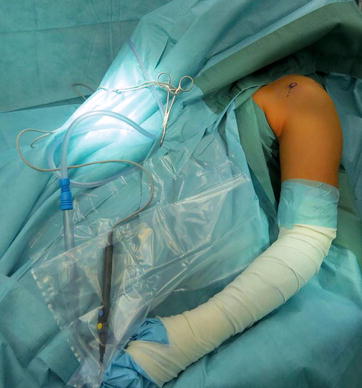
The patient is positioned in beach-chair position. The surgeon should check that no material will interfere with fluoroscopy. Frequently, the patient is positioned towards the injured side and the operating table is tilted towards the contralateral side to avoid the patient sliding down off the table. The arm is laid onto a splint and the elbow hangs freely (Fig. 4.1). The ventral and dorsal shoulder aspect as well as the sternoclavicular joint are cleaned and prepared for operation. The arm is freely movable and covered up to the biceps muscle.


Fig. 4.1
Arm position
4.5.2.2 Procedures
Regarding the anatomy, there are different options available. Bridging the acromioclavicular joint is possible; techniques that address the coracoid process are also available. Combinations of these options are possible as well.
Transarticular K-Wire Fixation
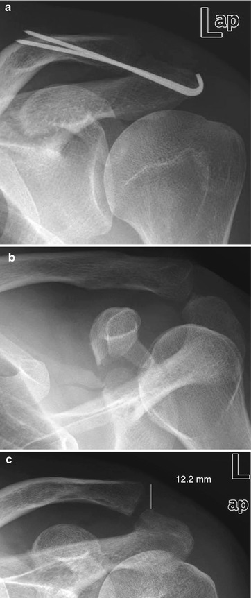
This technique was first described by Murray and Phemister. Using either a frontolateral approach parallel to the clavicle or a saber-type incision, preparation of the coracoclavicular and acromioclavicular ligaments as well as muscle insertions are possible. The insertion point for the K-wires can be set up. After reconstruction or resection of the ligaments and inspection and debridement of the discus has been performed, the clavicle is repositioned. The result is controlled by fluoroscopy before two K-wires (2 mm diameter) are drilled in from the lateral aspect of the acromion, aiming toward the cranial corticalis of the clavicle. Both wires should be positioned in parallel (Fig. 4.2a). Some authors prefer to use just one K-wire to reduce damage to the cartilage. It must be clearly stated that rotation stability is not achieved, which increases the risk of material loosening or even breakage.


Fig. 4.2


(a) Postoperative result. (b) Fracture of the coracoid process (oblique view). (c) Follow-up after metal removal (week 6), partial loss of reduction result is seen
Stay updated, free articles. Join our Telegram channel

Full access? Get Clinical Tree


