Fig. 29.1
Distribution of fracture frequency of fractures of the long bones in children (n = 678) (Taken from: Kindertraumatologie Marzi; Publisher: Steinkopff)
Statistically significant focuses are observed in three constellations. Firstly, fractures of the distal humerus in infants are mainly caused by playground accidents or at home. Secondly, fractures of the lower leg in older children and adolescents are associated with a high rate of traffic accidents. And thirdly, fractures of the distal forearm in all age groups are caused by sports-related traumas.
29.3 Post-traumatic Growth Disorders and Remodeling
The body is able to compensate remaining post-traumatic deformities to a certain extent by epiphyseal and periosteal correction mechanisms. The amount of the correction potential depends on the age of the child, the growth reserves of the corresponding physis, the location and direction of the dislocation, and the necessary correction direction. The periosteal correction is predominantly responsible for the correction of the ad latus displacement. Axis kinking in the diaphysis can also be compensated through periosteal correction mechanisms, because the concave side of the compression stimulates the bone regeneration, whereas bone is resorbed at the opposite convex side (Fig. 29.4). Simultaneously, the adjoint transversal standing physis adjusts orthogradely to the stress axis. Contractions can be compensated to a certain extent by growth stimulation within the fracture healing. A specific length adjustment is only possible in bone pairs such as the forearm. Rotation dislocations or post-traumatic elongations cannot be compensated or, if so, only poorly.
Elongations of the particular extremity must be expected because of corresponding growth stimulation that depends on the necessary remodeling. No functional consequences result at the upper extremity, so the spontaneous correction should be considered in the therapeutic strategies. At the lower extremity, larger dislocations should not be left to spontaneous correction to avoid leg distance differences with influence on the whole anatomical structure; thus, anatomic reposition should be the goal.
29.3.1 Growth Disorders
Stimulating and inhibiting growth disorders can be differentiated. Each fracture of a long bone leads to an excessive growth. The degree depends on the age of the child, the dislocation, the amount and the time of the reposition attempts, and the degree of the necessary remodeling. This leads to an elongation of the specific extremity, which is only relevant for the lower extremity concerning the biomechanical or anatomical structure of the spine. Therefore, regular clinical controls are necessary up to 2 years after the trauma. Partial growth stimulation is uncommon. It only plays a role in fractures of the proximal tibia with resulting increasing valgus or in condyle radial fractures with resulting increasing varus.
Injuries to the physis, directly or indirectly by vessel lesions, can cause early physis closure. A full closure leads to a shortening of the corresponding extremity. Growth disorders with increasing deformity of the axis are caused by incomplete closures. The degree depends on the age and the maturity of the child, the skeleton’s location, the proximity to the physis, and the degree of the dislocation. The occurrence of growth disorders cannot be avoided specifically. Ideal conditions, provided by correct anatomic reposition and avoidance of iatrogenic physis injuries, can only affect the appearance of growth disorders positively. In addition to clinical controls of the axis and length proportions in side comparison, an X-ray dense line, called the Harris line, which usually runs parallel to the physis, can provide a hint of partial growth disorders.
29.4 Characteristics of Injuries to the Immature Skeleton
29.4.1 Fracture Pattern
The specialties of the child’s skeleton with the physis, the strong ligaments, and the thick periost as well as the high elasticity of the bone leads to a stereotypic fracture pattern differing from that of adults. Therefore, special fracture forms exist for the child:
Bulge fracture: This fracture occurs in the area of the metaphysis, where the bone offers the highest porosity. It can be found in young children and it is called a stable and uncomplicated fracture. Usually, a therapeutic and short immobilization is sufficient for pain relief.
Bowing fracture: Because of the high elasticity of the child’s bone, a deformity occurs without a visible fracture. It can be apportioned functionally to the greenstick fractures.
Greenstick fractures: As a result of bowing, at the border of elasticity a greenstick fracture occurs. The trauma’s energy is not sufficient to break the bone completely. It is characterized by a full fracture of the side with the application of the force with intact opposite cortical. Because of the faster fracture healing and callus building of the opposite side, the danger of refracture exists in the diaphysis if fracture healing is absent in the area of the fracture gap.
Stress fractures: At the beginning of walking, an unused stress can cause an overload reaction of the child’s bones (toddler’s fracture). It mostly occurs at the tibia, the fibula, and the tarsal bones and cannot usually be seen in the first X-rays but impresses in the course by callus building.
29.5 Fracture Classification According to Salter and Harris (Figs. 29.2 and 29.17)
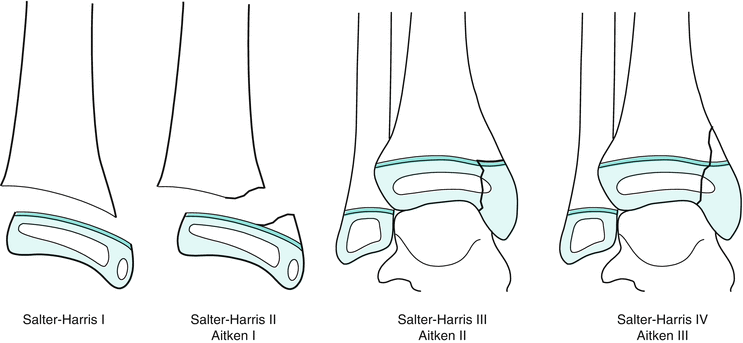
Fig. 29.2
Classification of the growth plate fractures according to Salter und Harris: Salter–Harris I: loosening of the growth plate; Salter–Harris II: growth plate loosening with metaphyseal wedge; Salter–Harris III: epiphyseal fracture; Salter–Harris IV: epiphyseal fracture with metaphyseal participation (Taken from: Kindertraumatologie Marzi; Publisher: Steinkopff)
Salter and Harris I: This refers to epiphysiolysis, usually as a result of shear injuries. If the periosteum remains intact, these fractures can appear almost undislocated and may be hardly recognizable radiologically. Clinically, local pressure pain and swelling are present solely over the concerned growth joint.
Salter and Harris II: If torsion forces also appear, a metaphysis wedge can break out next to the physis loosening, which is also called a Thurston-Holland fragment. An open reduction can become necessary in the affected periosteum.
Salter and Harris III: Here, the fracture line passes through the epiphysis into the joint.
Salter and Harris IV: The fracture passes through metaphysis and epiphysis.
Salter and Harris V: This describes an injury of the growth joint, caused by axial compression, without primary lesions visible in the X-ray. It presents primarily as a bruise or distortion and appears over the course of time as a growth disorder.
During adolescence, there is a slow closure of the physis, which leads to a changed fracture course. These fractures are generalized under the term “transitional fractures.” This fracture is most often seen in the distal tibia, which is described in the following example. It can, however, also appear at other localizations (Fig. 29.3). The affecting power deflects to the joint by the already ossified part of the physis, so that a more or less large ventrolateral epiphyseal fragment results as an ossified syndesmosis rupture, a so-called “two-plane fracture,” according to the size of the already resulting physis closure.
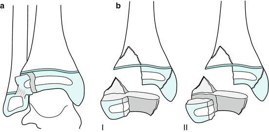
Fig. 29.3
Transition fractures of the distal tibia: (a) Two-plane fracture/Tilleaux fracture: epiphyseal fracture in already-beginning epiphyseal fusion; (b) Triplane fracture: epiphyseal fracture in already-beginning epiphyseal fusion with dorsal metaphyseal (triplane I) or epimetaphyseal wedge (triplane II) (Taken from: Kindertraumatologie Marzi; Publisher: Steinkopff)
Additional torsion powers can lead to a breakout of a dorsal additional fragment, corresponding to a Volkmann triangle, the so-called “tri-plane fracture.” If the metaphyseal fracture line ends in the joints, we speak of a “tri-plane-I fracture”; if it runs through the meta- and epiphysis, it is a “tri-plane-II fracture.” In comparison to fractures with a wide-open physis, the fracture lines often run obliquely. The Salter injury usually runs perpendicular to the physis, and mostly lies within the main burden of the joint zone. The fracture line of the Salter injury runs eccentrically far outside the stress-bearing area. Because of the low growth potential of these children, the reconstruction of the joint surface is the primary aim in these injuries, while relevant growth disorders are not to be expected at this age.
29.6 Diagnostics
29.6.1 Clinical Examination
The clinical investigation first covers the anamnesis, which identifies the localization of the injury, the energy of the trauma, and the relation of the trauma to possible child abuse or pathological fractures. After a thorough inspection, a careful palpation follows. Young patients should be informed about and provided with explanations of procedures. An examination of the peripheral blood circulation, motor function, and sensitivity should be performed in all cases. Expanded functional tests are mostly unnecessary because of lack of consequence in the acute stage and are potentially painful for the child.
29.6.2 Diagnostic Imaging
The X-ray diagnostics represent a special challenge. Radiation exposure should be minimized and all technical means should be used to reduce the radiation dose. Because of the large portion of X-ray-permeable cartilage tissue and the characteristics of the still-growing skeleton (e.g., the occurrence of ossification centers), good knowledge of age-dependent diagnostic findings is critical for the examiner. The frequently recommended comparative picture of the opposite side in the diagnosis of fresh injuries does not replace this knowledge and may be inefficient and unnecessary. If an operative indication is already provided by the first picture, a second plane can be avoided for the protection of the child. This can be accomplished under anesthesia. For all other fractures, the X-ray in two planes (anteroposterior, or a.p., and laterally) with illustration of the adjoining joint is standard. Injury-centered pictures should be aimed for and overview pictures in two planes should be avoided for the exact evaluation. In special cases such as transitional fractures, additional diagonal pictures are helpful. Due to the X-ray-permeable cartilage tissue, some injuries can only be recognized by an extension of the growth joint or a dislocation of the adjoining bone.
Sonography is available as a preserving procedure in the evaluation of X-ray-permeable structures such as tendons, ligaments, and non-ossificating skeleton portions. Fractures can also be diagnosed directly and indirectly by proof of a subperiosteal hematoma or an accompanying hemarthrosis.
Computed tomography, primarily as a multilayer CT, applies the diagnostics of the polytraumatized child as well as of complex fractures of the spine, the basin, or the foot root with the possibility of low-dose programs. MRI offers a large range of application possibilities. It allows the demonstration of nonossifying skeleton parts, soft tissues, ligaments, and musculature. It is used in the diagnosis of occult fractures, of spine injuries, and of post-traumatic complications of joint and physis injuries, such as the detection of growth disturbances, osteonecrosis, or cartilage flakes. The missing radiation exposure is a further advantage. However, it is still an expensive diagnostic and requires long investigation periods, and often requires sedation or anesthesia in small children.
Other imaging procedures such as scintigraphy, angiography, or arthrography are not applied in routine diagnostics and are reserved for special indications.
29.7 Treatment Options
The aim of therapy is to achieve fast fracture healing without the occurrence of complications, taking into account socioeconomic aspects. Special needs and wishes can be considered with the knowledge of the growth prognosis and complication possibilities. This is mainly relevant for the shaft; the closer the fracture is located to the joint, the greater the extent that the therapy is determined by the fracture. Adequate pain therapy should always be part of the primary therapy. Therefore, it may be necessary to perform splinting for immobilization and/or medicinal pain therapy before making a diagnosis.
29.7.1 Nonsurgical Treatment
In most cases, injuries of the child can be treated adequately by conservative techniques. Usually, an immobilization (e.g., with a cast) is sufficient. At certain localizations, the immobilization in small children takes place with special bandages (e.g., Desault’s bandage at the proximal humerus, the backpack bandage at the clavicle, or the fist bandage at the phalanxes). At certain localizations, secondary dislocations can be prevented and easy malpositions can be corrected by reduction bandages such as cast wedging, extension treatment, or the Cuff ‘n collar.
29.7.1.1 Cast Wedging
Axis deviations in the frontal and sagittal plane of the shaft zone can be corrected by cast wedging in a circular cast. This is primarily indicated in distal forearm fractures and tibia shaft fractures with a malposition in the frontal and/or sagittal plane. It is usually performed 8 days after primary cast immobilization. The primary swelling should have decreased by that point so that a secondary dislocation is not to be expected. A relative stability is given by the already-generated callus, which reduces the pain, with, however, still-existing plastic deformability, so as to accomplish a correction of the malposition.
29.7.1.2 Extension
The extension therapy in form of a strip extension only plays a role in the treatment of femur shaft fractures in infants. Disadvantages are the usually long stationary stay as well as the missing possibility to control the position of the fracture during the therapy. Other extension measures (e.g., “the hanging cast”) in the treatment of the humerus fracture no longer belong among the proven therapy methods.
29.7.1.3 Cuff ’n Collar (Blount Sling)
This provides a dynamic redress of a slightly in extension dislocated humerus fracture. By the adjustment of a bandage that gradually brings the elbow into the swelling-conditioned maximum pointed angle position, it leads to a slow correction of the malposition.
29.7.2 Surgical Treatment
All completely dislocated or unstable fractures that cannot be turned into a stable fracture by closed reduction are an indication for operative therapy. The aim is to achieve a stable situation to avoid further reductions or changes in therapy. A movement and charge stability should be achieved if possible. Furthermore, factors such as effort (e.g., secondary removal of metal), costs, available resources, and personal experience play a role in the choice of the osteosynthesis method.
29.7.2.1 Reduction
Dislocations that will not be corrected in the course of the spontaneous correction should be reduced. In order to avoid unnecessary pain and fear, this should be performed under general anesthesia and in operation standby in order to be able to proceed openly in case of reduction barriers and to be able to perform a stable osteosynthesis if there is danger of redislocation. A primarily open reduction should be performed in any joint fractures with a dislocation >2 mm, defect fractures, and partial open fractures. Generally, the first reduction should also be the last and final. In fractures of the phalanxes with significant dislocation, a reduction with local anesthesia can be performed.
29.7.3 Surgical Treatment Concepts
29.7.3.1 Kirschner Wire Osteosynthesis
This is indicated in metaphyseal fractures, including epiphysiolysis of the long tube bones, as well as in hand and foot fractures. In small children or in small fracture fragments, it can also be used in epiphyseal fractures. It can be placed minimally invasively, percutaneously, and the metal removal can usually be performed ambulatory without anesthesia. However, an additional cast immobilization is necessary for the protection of the stability.
29.7.3.2 Screw Osteosynthesis
This is especially indicated in joint fractures, inasmuch as a compression of the fracture gap can be caused, but also in epiphysiolysis with metaphyseal wedge. In principle, it is movement-stable, nevertheless, an additional protective immobilization with a cast is necessary. It can be used minimally invasively by closed reduction and by an open procedure. Therefore, cannulated, self-cutting cancellous bone screws are particularly suitable.
29.7.3.3 Tension Banding Osteosynthesis
This is indicated anywhere strong muscle or ligament originates. It can only be placed by an open procedure. Examples include patella transverse fracture, fracture of the olecranon, and lateral fracture of the clavicle.
29.7.3.4 Elastic Stable Intramedullary Nailing (ESIN)
This is a minimally invasive, movement and partially load-stable procedure in the treatment of dia- and metaphyseal shaft fractures. It’s basis is a three-point support of two inserted pretwisted flex, titanium nails within a tube bone which are inserted in opposite directions. The ideal fracture is a diaphyseal transverse fracture, but diagonal and spiral fractures can also be supplied by ESIN under adherence to basic biomechanical principles.
29.7.3.5 External Fixation
After ESIN, the external fixation represents the second most frequent method for the treatment of shaft fractures. It is mainly indicated in unstable long diagonal or multi-fragment fractures as well as in expanded tissue damage and for the fast treatment of the polytraumatized child.
29.7.3.6 Intramedullary Nailing
This is indicated in all tube bones with joints that have already been closed or with beginning joint closure, or in obese children. There is danger of damaging the physis directly or indirectly by damaging the supplying vessels (proximal femur) if the physis are still open. The procedure corresponds to the adult traumatology.
29.7.3.7 Plate Osteosynthesis
Plate osteosynthesis has been increasingly replaced by alternative methods like the ESIN in the treatment of shaft fractures. Usually, an open procedure with a large amount of tissue damage and cosmetically impairing scar formation is necessary. It remains subject to individual special indications like fractures of the phalanxes, of the hand and the foot, calcaneus fractures, and re-fractures of femur and tibia.
29.8 Special Fractures Types, Anatomical Distinctions
29.8.1 Clavicle
Clavicle fractures, accounting for 5–15 % of all fractures, children and adults, belong to the most frequent fractures in the infancy, mostly below the 10th year of life. They usually appear as birth trauma fracture or by direct impact. The fracture is often undislocated in infants because of the thick periosteal tube and leads to a fast fracture healing. Fractures are differentiated into the medial, middle, and lateral third; those in the middle third which occur most often. The therapy usually can be performed conservatively by immobilization in a cotton bandage or by backpack bandage. Consolidation control with still-open growth joints is done clinically by pressure indolent fracture callus and normalization of the function after approximately 3 weeks. Radiological position control is not necessary in that case. Shortenings or side-to-side malpositions will usually be corrected by the reduction within a year. In adolescents, the extent of the spontaneous correction is not sufficient for a complete remodeling. Pronounced malpositions and shortenings should be avoided in this case. In some cases, the danger of skin perforation exists; here, open reduction and osteosynthesis should be accomplished. In addition to the well-known method of plate osteosynthesis, intramedullary splinting by ESIN is a possible alternative.
29.8.2 Upper Extremity
29.8.2.1 Humerus
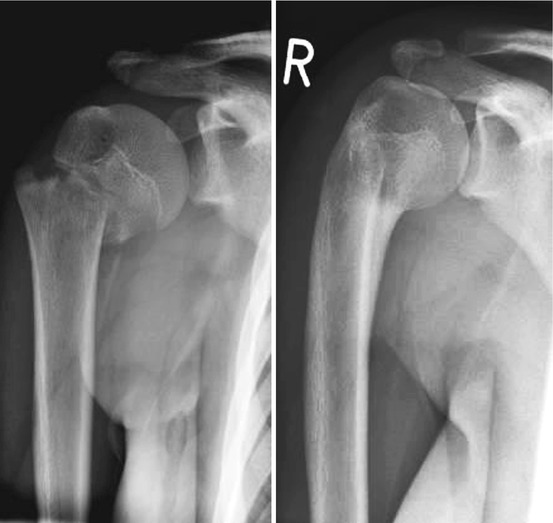
Proximal Humerus
In the majority of cases, fractures of the proximal humerus mostly concern subcapital fractures followed by epiphysiolysis, with or without a metaphyseal wedge (Fig. 29.4). Epiphyseal injuries are rare. The age peak is located between the 11th and 12th year. Because of the large growth potential of the proximal epiphysis joint, relatively large malpositions can be tolerated and left to spontaneous correction. Below the 10th year varus, anteversion and retroversion dislocation up to 50° and a valgus dislocation up to 10° serve as tolerance limits; in children older than 10 years, 20° and 10°, respectively. Conservative therapy with an immobilization in a Gilchrist or Desault bandage for 3–4 weeks is primary treatment. If the correction limits are exceeded, a reduction needs to be performed, usually in a closed manner. The first-choice osteosynthesis is the movement-stable retrograde ESIN nailing. Alternatively, a percutaneous K-wire osteosynthesis can be performed, which however, requires an additional immobilization, which is not always simple.