Fig. 75.1
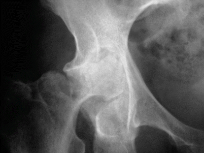
Plain radiograph of the hip showing a subchondral insufficiency fracture (two arrows)
Osteomalacia and osteoporosis are precursors of this problem. Analgesic use masks the pain of stress fracture and allows further weight bearing and destruction of bone.
Clinical Features
Epidemiology
Most common in middle-aged and older women.
Most patients have skeletal fragility.
Sites of Involvement
Femoral head, humeral head, pubis, pubic ramus, and the knee joint
Clinical Symptoms and Signs
Patients present with joint pain.
Analgesia allows weight bearing or use of the joint. This perpetuates a cycle of continued weight bearing and joint destruction (Fig. 75.2).

Fig. 75.2
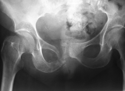
Plain radiograph of the hip in the same patient 6 months later showing destruction of much of the femoral head
Image Diagnosis
Radiographic Features
In the early phases, a subchondral insufficiency fracture can be noted in the femoral head.
Later portions of the femoral head disappear over weeks to months.
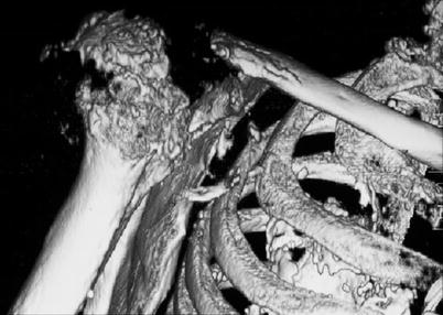
In the final stages, most of the femoral head or humeral head have disappeared.
In the knee, there is considerable discontinuity of the articular surface.
Traumatic osteolysis also occurs in the pubis (Fig. 75.3) and humerus (Fig. 75.4)

Fig. 75.3
Plain radiograph of the pubis showing pubic osteolysis with destruction of the right pubic ramus and ischium

Fig. 75.4
Reconstructive CT image of the rapidly destructive shoulder arthropathy. Almost the entire humeral head has been fragmented
Stay updated, free articles. Join our Telegram channel

Full access? Get Clinical Tree


