Fig. 72.1
Erdheim-Chester disease of the distal femur. There is a poorly defined zone of increased radiodensity with cortical thickening. This change is identical to chronic osteomyelitis
The cortices are also thickened due to periosteal new bone deposition.
Skeletal involvement is multifocal and is usually symmetrical (Fig. 72.2).
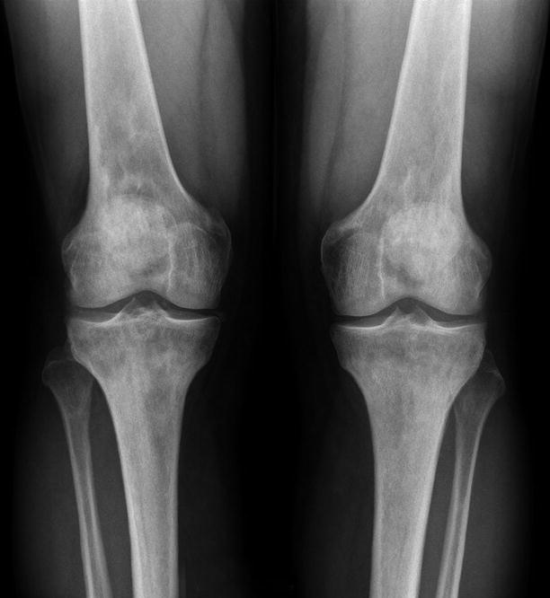
Fig. 72.2
Erdheim-Chester disease showing equal involvement of both femurs and both tibias
Bone scan shows widespread symmetrical lesions (Fig. 72.3).
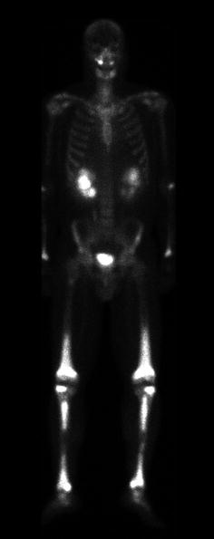
Fig. 72.3
Bone scan of Erdheim-Chester disease showing diffuse symmetrical uptake in the lower extremities
Image Differential Diagnosis
Chronic osteomyelitis
Not multifocal
Metastatic carcinoma
Usually a history of a primary neoplasm
Related posts:

Stay updated, free articles. Join our Telegram channel

Full access? Get Clinical Tree


