Fig. 54.1
(a, b) OFD. Anteroposterior and lateral radiographs show a typical case with bowing deformity of the tibia
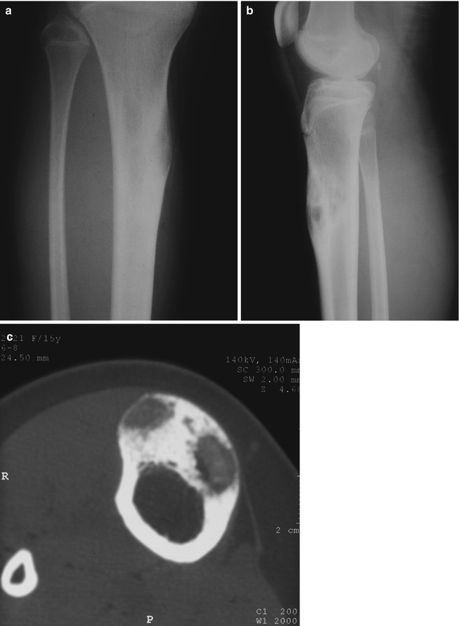
Fig. 54.2
(a, b) OFD. Plain x-ray of the tibia showing the fusiform cortical expansion of the anterior cortex. (c) CT scan shows multilocular defect in thickened anteromedial cortex of the tibia
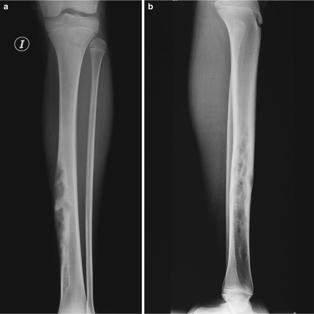
Fig. 54.3
(a, b) Plain radiographs illustrating a large and elongated lesion with multiple cortical lucencies of the anterior cortex of the tibia
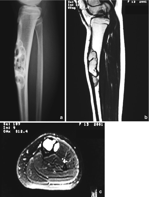
Fig. 54.4


(a) Lateral x-ray showing an expansile lucent lesion surrounded by sclerosis and thinning of the anterior aspect of the tibia. (b and c) Sagittal and axial MRI of the lesion
Related posts:

Stay updated, free articles. Join our Telegram channel
Full access? Get Clinical Tree
