Fig. 58.1
Radiograph of a solitary Langerhans cell histiocytosis (eosinophilic granuloma) in the femoral diaphysis of a young child. Lytic lesion with extensive reactive sclerosis and lamellar periosteal reaction. Radiologic differential diagnosis with Ewing sarcoma and osteomyelitis
Fig. 58.2
(a) Radiograph of the spine of another young child showing a vertebra plana, flattening of the vertebral body, common occurrence in LCH of the vertebra. (b) MRI of same case of previous figure, better demonstrating the flattened vertebra. (c) Needle biopsy of the lesion in previous figure retrieved some hemorrhagic material. Processing of the blood clots disclosed small fragments sufficient for the diagnosis of LCH (d)
Fig. 58.3
(a) Radiograph of LCH affecting the lumbar spine. Lytic lesion in the body and pedicles of a vertebra can be seen. Vertebra shape is maintained. (b) MPR CT of the same case as previous figure. Although vertebral height is maintained, a lytic lesion extends to its posterior elements. (c) Same case as previous figure. MRI demonstrates better the extension of the lesion
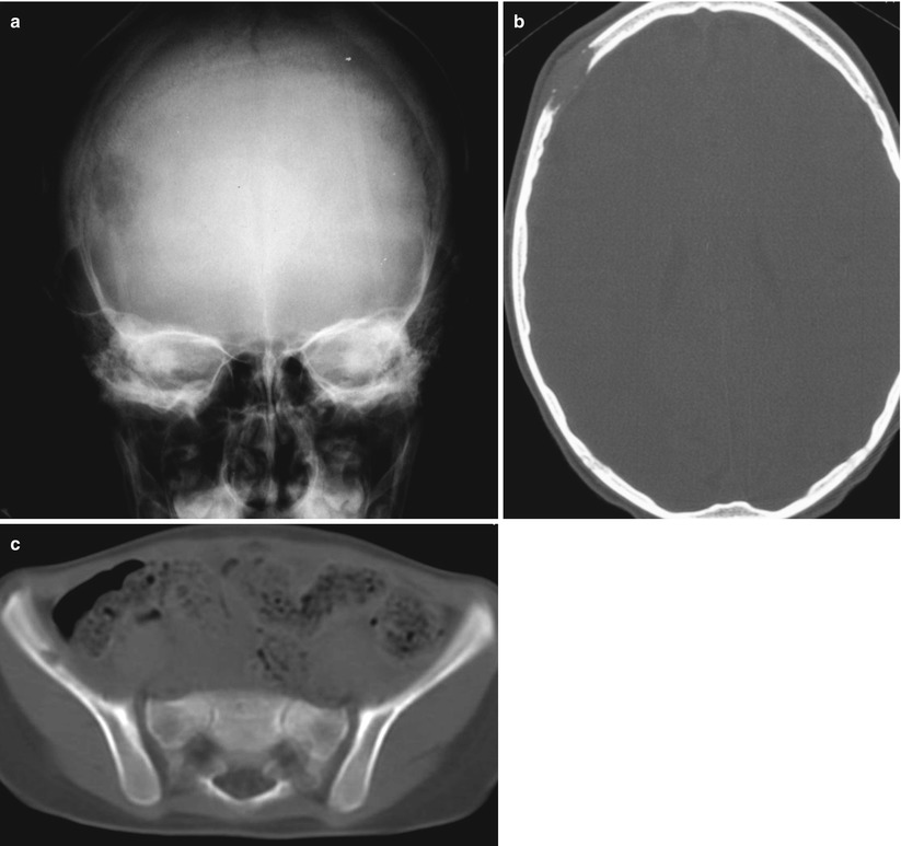
Fig. 58.4
(a) Radiograph of a skull lytic geographic lesion of LCH. (b) CT scan of the same case of (a). Characteristic beveled destruction of the two cranial cortices, responsible for the “hole-in-hole” aspect of radiographs. (c) CT scan of the pelvis of the same patient of (a). Secondary focus of LCH
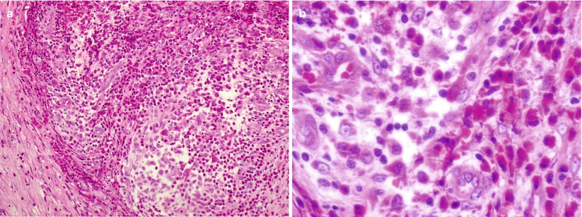
Fig. 58.5
(a) Histologic low-power view of an LCH lesion. Dense proliferation of histiocytes with abundant eosinophils, surrounded by fibrous tissue, can be seen even in this magnification. (b) Higher magnification of previous histologic field depicting histiocyte and eosinophil proliferation
Fig. 58.6
(a) Histologic low-power view of another LCH lesion, in this case with dense, almost exclusive histiocyte proliferation. (b) Higher magnification of same case of previous figure. Indented nuclei of the histiocytes can be seen. Eosinophils and other inflammatory cells can also be identified among the histiocytes