Fig. 17.1
Radiograph shows multiple enchondromas involving whole digits and metacarpal bones leading to extreme deformity
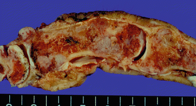
Fig. 17.2
Gross specimen, as illustrated in Fig. 17.1, shows numerous grayish-blue cartilage masses within the marrow cavity of the phalangeal bones
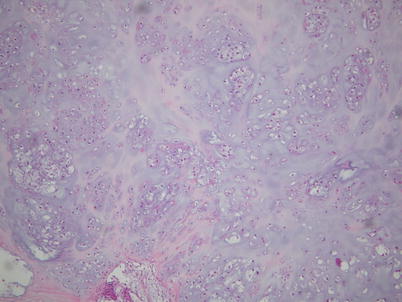
Fig. 17.3
Low-power microscopic view shows lobulated cartilage masses and rich cellularity
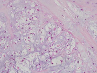
Fig. 17.4
At the periphery of the cartilage lobule, there are enlarged nuclei and binucleation of the chondrocytes, associated with myxoid matrix degeneration
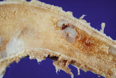
Fig. 17.5
Specimen photography showing several medullar enchondromas in a same bone
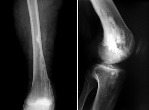
Fig. 17.6
AP and lateral radiographs of a case of Ollier’s disease showing characteristic finger-like longitudinal chondromatous proliferations
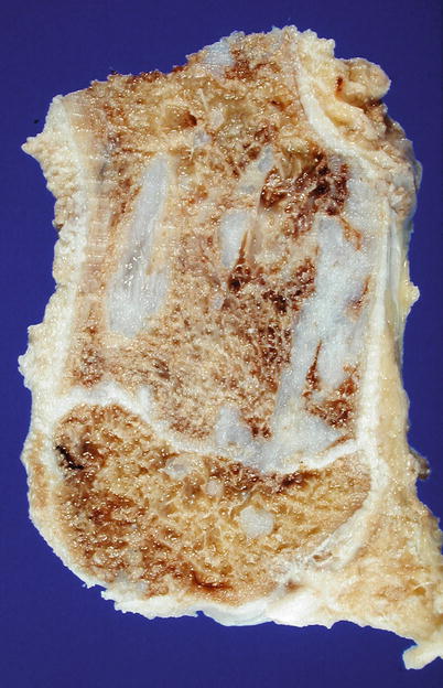
Fig. 17.7
Macroscopic demonstration of fingerlike chondromatous proliferations in Ollier’s disease
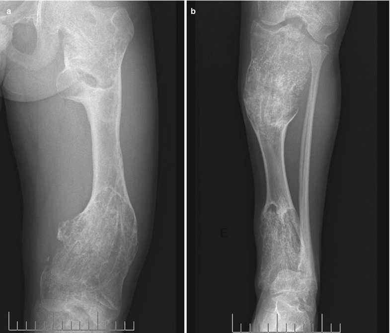
Fig. 17.8


Enchondromatosis (Ollier’s disease). Radiographs of femur (a) and tibia (b) showing multiple deformities caused by large enchondroma lesions in metaphyseal location. Lesions present characteristic elongated ridges and disseminated calcified spots
Related posts:

Stay updated, free articles. Join our Telegram channel
Full access? Get Clinical Tree
