Fig. 38.1
Benign notochordal cell tumor involving the S3 of the sacrum in a 31-year-old man. Sagittal (a) and axial (b) computed tomographic scans show a slightly sclerotic mass in midline of the S3 in the sacrum. Sagittal (c) and axial (d) T1-weighted magnetic resonance images demonstrate low signal intensity, without significantly increased intensity in gadolinium-DTPA-enhanced T1-weighted image (e). Sagittal (f) and axial (g) T2-weighted magnetic resonance images show heterogeneously high signal intensities
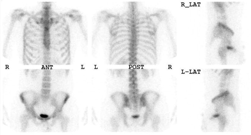
Fig. 38.2
No abnormal uptake in bone isotope scan
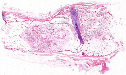
Fig. 38.3
Low-power scanning view of a benign notochordal cell tumor showing a relatively well-circumscribed medullary nodule containing slightly sclerotic bony trabeculae (Case by courtesy of Doctor Takehiko Yamaguchi, Tochigi, Japan)
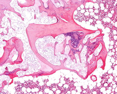
Fig. 38.4
Benign notochordal cell tumor permeating the intertrabecular spaces with various degree of osteosclerotic change (Case by courtesy of Doctor Takehiko Yamaguchi, Tochigi, Japan)
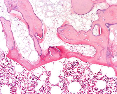
Fig. 38.5
Proliferation of bland-looking tumor cells resembling fatty marrow between bony trabeculae (Case by courtesy of Doctor Takehiko Yamaguchi, Tochigi, Japan)
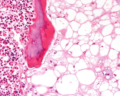
Fig. 38.6
Faintly eosinophilic cytoplasm with prominent vacuolization, mimicking mature adipocytes (Case by courtesy of Doctor Takehiko Yamaguchi, Tochigi, Japan)
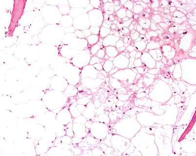
Fig. 38.7
Benign notochordal cell tumor (center to right) continuously merging into fatty marrow (left) (Case by courtesy of Doctor Takehiko Yamaguchi, Tochigi, Japan)
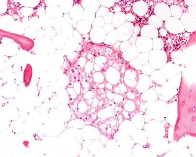
Fig. 38.8
Discretely isolated small foci of giant notochordal tumor in the fatty bone marrow (Case by courtesy of Doctor Takehiko Yamaguchi, Tochigi, Japan)
Fig. 38.9
Inconspicuous neoplastic cells between bony trabeculae (a) are positive for keratin (CAM5.2) in immunostaining (b)
Recommended Reading
Adamek D, Malec M, Grabska N, Krygowska-Wajs A, Galazka K. Ecchordosis physaliphora – a case report and a review of notochord-derived lesions. Neurol Neurochir Pol. 2011;45:169–73.PubMed
Alkan O, Yildirim T, Kizilkilic O, Tan M, Cekinmez M. A case of ecchordosis physaliphora presenting with an intratumoral hemorrhage. Turk Neurosurg. 2009;19:293–6.PubMed
Amer HZ, Hameed M. Intraosseous benign notochordal cell tumor. Arch Pathol Lab Med. 2010;134:283–8.PubMed
Deshpande V, Nielsen GP, Rosenthal DI, Rosenberg AE. Intraosseous benign notochord cell tumors (BNCT): further evidence supporting a relationship to chordoma. Am J Surg Pathol. 2007;31:1573–7.CrossRefPubMed
Related posts:

Stay updated, free articles. Join our Telegram channel

Full access? Get Clinical Tree


